
Translate this page into:
Thalamic Hamartoma Presenting with Tremor
Address for correspondence: Dr. Niraj Kumar, Department of Neurology, All India Institute of Medical Sciences, Rishikesh, Uttarakhand, India. E-mail: drnirajkumarsingh@gmail.com
This is an open access journal, and articles are distributed under the terms of the Creative Commons Attribution-NonCommercial-ShareAlike 4.0 License, which allows others to remix, tweak, and build upon the work non-commercially, as long as appropriate credit is given and the new creations are licensed under the identical terms.
This article was originally published by Medknow Publications & Media Pvt Ltd and was migrated to Scientific Scholar after the change of Publisher.
A 15-year-old female, with noncontributory past medical and family history, presented with gradually progressive right upper-extremity action tremor of 8-year duration. Neurological examination revealed right upper-extremity postural and kinetic tremor [Video 1]. Laboratory workup revealed a normal complete blood count, serum electrolytes, creatinine, glucose, liver, and thyroid function test. Magnetic resonance imaging (MRI) brain showed left thalamic mass lesion with the periphery appearing hyperintense on T1 and hypointense on T2 and gradient sequences and cerebrospinal fluid-like signal intensity in the center. Moreover, minimal postcontrast enhancement [Figure 1]. A noncontrast computed tomography brain showed dense calcification involving the thalamic lesion. The radiological findings favored left thalamic hamartoma. The tremor was medication-refractory, and the family deferred neurosurgical therapy.
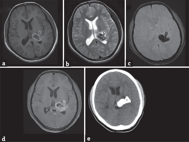
- Magnetic resonance imaging brain (a-d) shows a large lobulated lesion involving left thalamus with beaklike protrusion in the adjacent brain parenchyma. The periphery of the lesion appears hyperintense on T1-weighted image (a) and hypointense on T2-weighted and gradient recalled echo images (b and c) with minimal postcontrast enhancement (d). The center of the lesion shows cerebrospinal fluid-like signal intensity on all sequences. Non-contrast CT brain (e) shows dense calcification in the lesion
Neuronal hamartoma is a rare disorder, commonly arising in the hypothalamus.[1] It is more common in males, with precocious puberty, pressure effects, and gelastic epilepsy being usual presenting features.[1] Till date, only two cases with hamartoma originating in the thalamus have been reported.[12] While one case presented at the age of 8 with left upper-extremity tremor and weakness,[1] the other was born with severe macrocephaly and several congenital abnormalities including bilateral cleft lip and palate, right microphthalmos, left anophthalmos, and left foot equinovarus deformity with sudden death at the age of 7 years.[2] Our case had right upper-extremity action tremor with no cardiac, ocular, or skeletal abnormality. MRI brain in neuronal hamartoma may show calcification as was seen in our case.[1] Ischemic changes resulting from expanding hamartoma size may cause calcification.[13] Minimal contrast enhancement in hamartoma may result from vascular hyperplasia.[1]
Declaration of patient consent
The authors certify that they have obtained all appropriate patient consent forms. In the form, the patient has given her consent for her images and other clinical information to be reported in the journal. The patient understand that names and initials will not be published and due efforts will be made to conceal identity, but anonymity cannot be guaranteed.
Financial support and sponsorship
Nil.
Conflicts of interest
There are no conflicts of interest.
Video Available on: www.ruralneuropractice.com
REFERENCES
- Degenerative thalamic hamartoma: CT and MR imaging features. AJNR Am J Neuroradiol. 2004;25:766-8.
- [Google Scholar]
- Diencephalic neuronal hamartoma associated with congenital obstructive hydrocephalus, anophthalmia, cleft lip and palate and severe mental retardation: A possible new syndrome. Acta Neuropathol. 2000;99:685-90.
- [Google Scholar]
- Giant hypothalamic hamartoma with cystic change: Report of two cases and review of the literature. Neuroradiology. 2000;42:648-50.
- [Google Scholar]


