
Translate this page into:
Pie in the sky appearance – Unraveling rheumatic heart disease in a pediatric stroke
*Corresponding author: Arvinder Wander, Department of Pediatrics, All India Institute of Medical Sciences, Bathinda, Punjab, India. wander1686@gmail.com
-
Received: ,
Accepted: ,
How to cite this article: Singh R, Lakhanpal V, Dhoot SK, Peer S, Wander A. Pie in the sky appearance – Unraveling rheumatic heart disease in a pediatric stroke. J Neurosci Rural Pract. 2024;15:607-9. doi: 10.25259/JNRP_158_2024
Abstract
We present a very interesting case of pediatric stroke whereby the patient presented with a visual field defect with perimetry showing a “Pie in the Sky” appearance and ultimately diagnosed with rheumatic heart disease on stroke workup.
Keywords
Quadrantanopia
Rheumatic heart disease
Pediatric stroke
Perimetry
Magnetic resonance imaging

A 13-year-old female presented with a two-month history of difficulty in visualizing the upper portion of the television screen. Perimetry [Figure 1] revealed left homonymous superior quadrantanopia. Magnetic resonance imaging brain [Figures 2a-f] showed a chronic infarct in the right occipitotemporal gyrus, corresponding to the location of the optic radiation. A chronic lacunar infarct was also noted in the right hemi-thalamus and in the right cerebellar hemisphere. The presence of multiple infarcts raised the possibility of embolic etiology. Electrocardiography (ECG) was normal. A 2D echocardiography revealed moderate mitral stenosis with a thickened anterior mitral valve leaflet, suggestive of rheumatic heart disease (RHD). Further, stroke workup including testing for hypercoagulable states, vasculitis, and other embolic sources was negative. A 24 h Holter ECG monitoring was also normal with no evidence of cardiac arrhythmias.[1] The patient was initiated on penicillin prophylaxis and anti-coagulants.
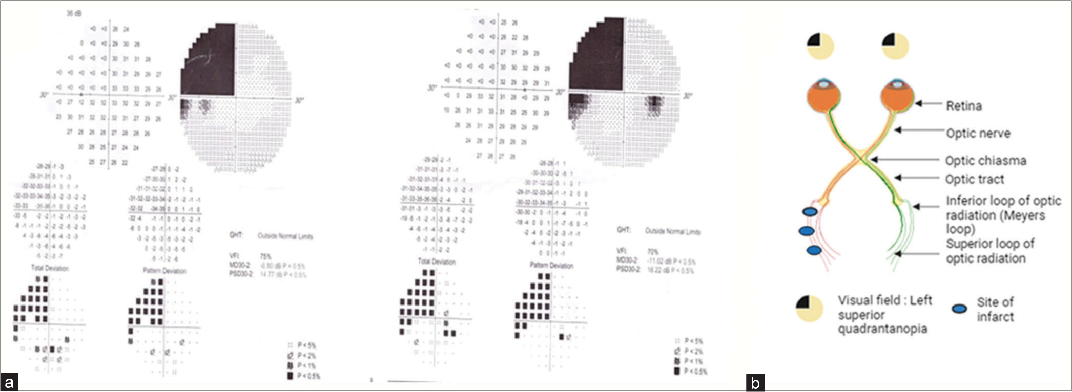
- (a) Perimetry showing left homonymous superior quadrantanopia, (“pie in the sky,” appearance) on Humphrey visual field 30-2. (b) Diagram showing optic pathway with lesion in inferior fibers of right optic radiation causing left homonymous superior quadrantanopia.
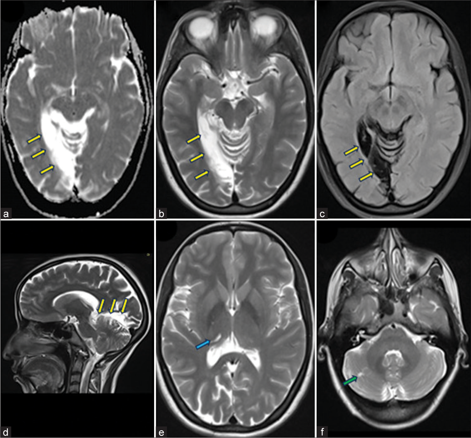
- (a-c) Axial and (d) Sagittal MRI brain showing chronic infarct in the right occipitotemporal gyrus (yellow arrows), corresponding to location of optic radiation. (e-f) Axial MRI brain showing a chronic lacunar infarct in right hemi-thalamus (blue arrow) and in right cerebellar hemisphere (green arrow). Presence of multiple infarcts raised the possibility of embolic etiology.
Homonymous superior quadrantanopia results from damage to the contralateral inferior fibers of the optic radiation (Meyer’s loop) or the inferior part of the occipital visual cortex.[2] The RHD patients have a significantly higher risk of stroke compared to the normal population.[3] This case exhibited the characteristic “pie in the sky” appearance due to infarct involving the inferior fibers of the optic radiation. A stroke workup in young patients with visual field defects is essential for diagnosing latent diseases, as demonstrated in this case of latent mitral valve disease leading to multiple cerebral emboli and resultant stroke.
Ethical approval
The Institutional Review Board approval is not required.
Declaration of patient consent
The authors certify that they have obtained all appropriate patient consent.
Conflicts of interest
There are no conflicts of interest.
Use of artificial intelligence (AI)-assisted technology for manuscript preparation
The authors confirm that there was no use of artificial intelligence (AI)-assisted technology for assisting in the writing or editing of the manuscript and no images were manipulated using AI.
Financial support and sponsorship
Nil.
References
- The significance of Holter electrocardiography in the etiological evaluation of transient ischemic stroke. Brain Circ. 2020;6:191-5.
- [CrossRef] [PubMed] [Google Scholar]
- The localizing value of a quadrantanopia. Arch Neurol. 1997;54:401-4.
- [CrossRef] [PubMed] [Google Scholar]
- Epidemiologic assessment of chronic atrial fibrillation and risk of stroke: The Framingham study. Neurology. 1978;28:973-7.
- [CrossRef] [PubMed] [Google Scholar]


