Translate this page into:
Marcus Gunn phenomenon with abducens palsy: As pseudo-false localizing signs
Address for correspondence: Dr. G Samson Sujit Kumar, Department of Neurosurgery, Narayana Medical College, Chintareddypalem, Nellore-524002, A.P., India, E-mail: gsskcmc@yahoo.com
This is an open-access article distributed under the terms of the Creative Commons Attribution-Noncommercial-Share Alike 3.0 Unported, which permits unrestricted use, distribution, and reproduction in any medium, provided the original work is properly cited.
This article was originally published by Medknow Publications and was migrated to Scientific Scholar after the change of Publisher.
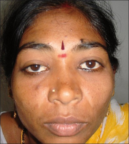
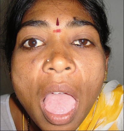
We report a case of a 25-year-old woman presented with history of headache and right upper limb focal sensory seizures of 3 months duration. On examination, her vitals were stable and she had bilateral papilledema. There were no focal motor or sensory deficits. She had mild ptosis and lateral rectus palsy of her right eye. Magnetic resonance imaging (MRI) of the brain showed an enhancing left parietal convexity dural-based lesion with perilesional edema and midline shift. On careful examination we found that on opening the mouth, ptosis in the right eye disappeared and there was widening of the palpebral fissure, a feature typical of Marcus Gunn phenomenon (Marcus Gunn jaw winking or trigemino-oculomotor synkineses) [Figures 1 and 2]. Forced abduction test did not reveal fibrosis of the right lateral rectus but from history this patient had partial ptosis and inability to abduct the right eye since birth. She underwent left parietal craniotomy and total excision of meningioma. We report this case to highlight the importance of detailed history, careful examination, and correct interpretation of neurologic signs. Apart from meningioma we make a collateral diagnosis of congenital Marcus Gunn phenomenon with a very rare association of lateral rectus palsy presenting in this case as pseudo false localizing signs (because these signs of ptosis and lateral rectus paresis, which are usually false localizing signs in a setting of raised intracranial pressure, are not actually due to the effect of meningioma and are present before the symptoms started).

- Clinical picture of the patient taken at rest showing right eye partial ptosis

- Clinical picture of the patient taken on opening the mouth showing widening of the right eye palpebral fi ssure
Marcus Gunn phenomenon was first described by ophthalmologist Marcus Gunn.[1] This condition presents in approximately 5% of neonates with congenital ptosis. It is associated with amblyopia (30%–60% of cases), anisometropia (5%–25% of cases), and strabismus (50%–60% of cases).[2] It is usually sporadic but an autosomal dominant variety with incomplete penetrance has also been reported.[3] Marcus Gunn phenomenon (Marcus Gunn jaw winking or trigemino-oculomotor synkineses) is a classical example of synkinesis in which 2 or more muscles that are independently innervated have either simultaneous or coordinated movements. Common physiologic examples of synkineses occur during sucking, chewing, or conjugate eye movements. Pathologic causes of synkinesis are either acquired (trauma, infection, surgical) or congenital. Examples of acquired synkineses include facial synkinesis (inverse Marcus Gunn phenomenon—closing of the palpebral fissure on opening the mouth; crocodile tears—tears from the eyes while eating), extraocular muscle synkinesis, and trigemino-abducens synkinesis. In these conditions, there is aberrant regeneration of the cranial nerves following inflammation (eg, facial nerve in Bell's palsy), surgical procedures, or trauma. Marcus Gunn phenomenon is an example of pathologic congenital synkinesis. The postulated mechanism is loss of inhibition of primitive reflexes along with their associated movements following congenital cranial nerve nuclear lesions in the brain stem.[4] Some authors suggest an abnormal connection between the motor branch of the trigeminal nerve and the branch of oculomotor nerve supplying the levator palpebri superioris.[56] Stimulation of trigeminal nerve by contraction of the pterygoid muscles results in abnormal excitation of the branch of oculomotor nerve that innervates the levator palpebri superioris. It could be of 2 types: external pterygoid–levator synkinesis where there is raising of upper eyelid on opening the mouth (more common type) and the internal pterygoid-levator synkinesis where there is raising of upper eyelid on clenching of the teeth. This phenomenon should be considered in the differential diagnosis of ptosis, especially in children. In our case, this phenomenon along with abducens palsy presents as a pseudo false localizing sign in a setting of raised intracranial pressure.
Source of Support: Nil
Conflict of Interest: None declared.
References
- Congenital ptosis with peculiar associated movements of the affected lid. Trans Ophthal Soc UK. 1883;3:283-7.
- [Google Scholar]
- Normal and abnormal development: Congenital deformities. In: System of Ophthalmology. Vol 3. St. Louis: CV Mosby; 1963. p. :900-5.
- [Google Scholar]


