Translate this page into:
Isolated Intracranial Myeloid Sarcoma Occurring as Relapse in Acute Myeloid Leukemia
Address for correspondence: Dr. Geetha Narayanan, Department of Medical Oncology, Regional Cancer Centre, Thiruvananthapuram - 695 011, Kerala, India. E-mail: geenarayanan@yahoo.com
This is an open access article distributed under the terms of the Creative Commons Attribution-NonCommercial-ShareAlike 3.0 License, which allows others to remix, tweak, and build upon the work non-commercially, as long as the author is credited and the new creations are licensed under the identical terms.
This article was originally published by Medknow Publications & Media Pvt Ltd and was migrated to Scientific Scholar after the change of Publisher.
Abstract
Myeloid sarcoma (MS) or chloroma is a rare extramedullary tumor composed of extramedullary proliferation of blasts of granulocytic, monocytic, erythroid, or megakaryocytic lineage occurring at sites outside the bone marrow. MS occurs in 2%–8% of patients with acute myeloid leukemia (AML), sometimes it occurs as the presenting manifestation of relapse in a patient in remission. We describe the case of a young male with AML in remission for 6 years presenting with central nervous system symptoms. Magnetic resonance imaging showed an extra-axial altered intensity lesion in the parasagittal parietal region, infiltrating anterosuperiorly into anterior falx, and posterosuperior aspect of the superior sagittal sinus. A biopsy from the lesion was diagnostic of MS which was positive for myeloperoxidase. He did not have any other sites of disease. He has received chemotherapy with FLAG (Fludarabine, Cytosine arabinoside) followed by cranial irradiation and is in complete remission.
Keywords
Intracranial
isolated
myeloid sarcoma
INTRODUCTION
Myeloid sarcoma (MS) or chloroma is a rare extramedullary tumor composed of an extramedullary proliferation of blasts of granulocytic, monocytic, erythroid, or megakaryocytic lineage occurring at sites outside the bone marrow. MS occurs in 2%-8% of patients with acute myeloid leukemia (AML). it can precede the diagnosis of AML, or it can occur concurrently with diagnosis of AML or it may occur during the course of AML. Sometimes, it occurs as the presenting manifestation of relapse in a patient in remission.[123] We describe the case of a young male with AML in remission for 6 years relapsing with an intracranial mass.
CASE REPORT
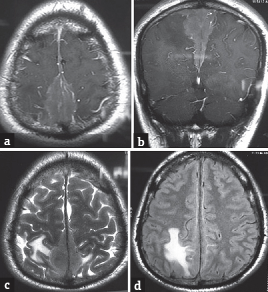
A 20-year-old male patient was diagnosed as AML M5a with hypodiploidy and treated with induction chemotherapy and three cycles of high-dose cytosine arabinoside. He was asymptomatic, in complete remission, and on follow-up for 6 years when presented with a headache for 5 months. The headache was insidious in onset, bifrontal and was associated with vomiting. He also had two episodes of seizures. He did not have any visual symptoms, facial asymmetry, nasal regurgitation, or gait disturbance. On examination, the patient was conscious, higher motor functions were normal, pupils were equal and reactive, and cranial nerves and fundi were normal. A magnetic resonance imaging showed an extra-axial altered intensity lesion measuring 42 mm × 37 mm in the parasagittal parietal region. The lesion was seen infiltrating anterosuperiorly along the interhemispheric dura into anterior falx and posterosuperior aspect of the superior sagittal sinus. The lesion was isointense on T1 and T2 showing diffusion restriction and enhances homogenously on contrast administration. Flair hyperintense was noted in the posterior parietal and superior frontal lobes along the lesion suggestive of edema. The picture was suggestive of leukemic infiltration causing pachymeningitis [Figure 1a-d].

- (a) Postcontrast T1-weighted magnetic resonance imaging brain showing homogenous enhancement. (b) Postcontrast T1-weighted magnetic resonance imaging brain coronal view. (c) T2-weighted magnetic resonance imaging showing isointense lesion with diffusion restriction and homogenous enhancement on contrast. (d) Magnetic resonance imaging showing flair hyperintensities suggestive of edema
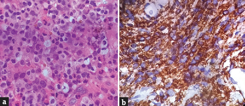
The patient underwent the right pericoronal parasagittal craniotomy and biopsy. The histopathological examination showed tissue infiltrated by atypical medium-sized cells with moderate cytoplasm and indented irregular nuclei. On immunohistochemistry (IHC), the abnormal cells were positive for CD68, CD34, CD117, CD11c, and myeloperoxidase (MPO) [Figure 2a and b]. The cells were negative for CD20, CD5, and CD3. A diagnosis of intracranial MS was made. His blood counts, bone marrow, and cerebrospinal fluid studies were normal. He received salvage chemotherapy with fludarabine and cytosine arabinoside (FLAG) for 3 cycles along with triple intrathecal chemotherapy followed by cranial irradiation (24Gy). Currently he is in complete remission and on follow up.

- (a) Tissue infiltrated by abnormal cell (H and E, ×100). (b) Tumor cells positive for myeloperoxidase (IHC, ×100)
DISCUSSION
Apart from AML, MS can occur in chronic myeloid leukemia and other chronic myeloproliferative disorders. The common sites for MS are bone, soft tissue, lymph node, and skin. Rarely, they can involve the central nervous system (CNS), gastrointestinal tract, genitourinary tract, breast, and testes.[1234]
MS is commonly seen in children and young adults and has a male predilection.[2] MS of CNS is a rare presentation of AML and may involve the subperiosteum, dura mater, and occasionally the brain parenchyma. Leukemic cells invade the CNS through the bone marrow of the adjacent cranium, vertebrae, or orbital bones.[56] Clinically, it may present as a meningeal disease (carcinomatous meningitis) or intravascular tumor aggregates in the brain (carcinomatous encephalitis) or as local tumor mass of MS.[7] The prevalence of MS in AML was 9.7%, but MS of the CNS was only 0.4% in a population-based study.[8]
In their review on the radiology of 24 intracranial MS, 48% presented as intraparenchymal tumor mass.[7] Radiologically, intracranial MS presents as an extra-axial hyperdense mass on noncontrast computed tomography scan and thus mimicks a meningioma, lymphoma, or metastasis.[7] Another pattern of the presentation was patchy leptomeningeal enhancement, mimicking leptomeningeal carcinomatosis, meningitis, neurosarcoidosis, dural sinus thrombosis, etc. Vasogenic edema was present in 45% of cases, and destructive bony changes are uncommon.[7] This patient presented with isolated MS of the brain as the first sign of relapse after 6 years of remission.
MS is classified into granulocytic, monoblastic, or myelomonocytic based on the predominant cell type. Microscopically, the characteristic pattern is Indian file pattern, and the MIB index is usually high.[24] IHC and immunophenotyping (IPT) are essential for a correct diagnosis. MS has IPT features identical to leukemic cells, the most common positive markers are CD68, MPO, CD117, CD99, CD34, Tdt, CD56, CD61, CD30, glycophorin, and CD4. MS is associated with chromosomal abnormalities such as AML and those with t(8:21) translocation have a predilection for MS of the CNS.[9] The present case was positive for MPO, CD68, CD117, and CD11c.
The currently recommended treatment for MS is combination chemotherapy similar to AML. There is no prognostic clinical or pathologic features, however, in patients who undergo allogeneic bone marrow transplantation, the survival is better.[210] The outcome of 23 patients with MS was compared with AML, and the event-free survival was found to be longer in patients with isolated MS.[1] Our patient received salvage chemotherapy with FLAG followed by cranial irradiation and is on follow up.
CONCLUSION
AML relapsing as an isolated intracranial MS is rare. A high index of suspicion for extramedullary disease should be kept in patients with AML on follow-up who present with CNS symptoms. This is important for early diagnosis and salvage treatment.
Financial support and sponsorship
Nil.
Conflicts of interest
There are no conflicts of interest.
REFERENCES
- Myeloid sarcoma is associated with superior event-free survival and overall survival compared with acute myeloid leukemia. Cancer. 2008;113:1370-8.
- [Google Scholar]
- Myeloid sarcoma: Clinico-pathologic, phenotypic and cytogenetic analysis of 92 adult patients. Leukemia. 2007;21:340-50.
- [Google Scholar]
- Granulocytic sarcoma: A clinicopathologic study of 61 biopsied cases. Cancer. 1981;48:1426-37.
- [Google Scholar]
- Prognostic factors of treatment outcomes in patients with granulocytic sarcoma. Acta Haematol. 2009;122:238-46.
- [Google Scholar]
- Outcome in patients with nonleukemic granulocytic sarcoma treated with chemotherapy with or without radiotherapy. Leukemia. 2003;17:1100-3.
- [Google Scholar]
- Myeloid sarcoma with multiple lesions of the central nervous system in a patient without leukemia. Case report. J Neurosurg. 2006;105:916-9.
- [Google Scholar]
- Intracranial CNS manifestations of myeloid sarcoma in patients with acute myeloid leukemia: Review of the literature and three case reports from the author's institution. J Clin Med. 2015;4:1102-12.
- [Google Scholar]
- Extramedullary Leukemia and Myeloid Sarcoma in AML: New Clues to an Old Issue. Results of a Retrospective Population-Based Registry-Study of 2261 Patients; Annals of 53rd American Society of Hematology Annual Meeting and Exposition; 2011, San Diego, CA, USA; 10-13 December 2011
- Isolated recurrence of granulocytic sarcoma manifesting as extra- and intracranial masses – Case report. Neurol Med Chir (Tokyo). 2004;44:311-6.
- [Google Scholar]
- Clinico-pathological characteristics of myeloid sarcoma at diagnosis and during follow-up: Report of 12 cases from a single institution. Leuk Res. 2004;28:1165-9.
- [Google Scholar]


