Translate this page into:
Histologically Confirmed Intracranial Tumors Managed at Enugu, Nigeria
Address for correspondence: Dr. Chika Anele Ndubuisi, Memfys Hospital for Neurosurgery, P.O.Box 2292, Enugu, Nigeria. E-mail: chikandu@yahoo.com
This is an open access article distributed under the terms of the Creative Commons Attribution-NonCommercial-ShareAlike 3.0 License, which allows others to remix, tweak, and build upon the work non-commercially, as long as the author is credited and the new creations are licensed under the identical terms.
This article was originally published by Medknow Publications & Media Pvt Ltd and was migrated to Scientific Scholar after the change of Publisher.
Abstract
Background:
There is controversy about the global distribution of intracranial tumors (ICTs). The previous reports from Africa suggested low frequency and different pattern of distribution of brain tumors from what obtains in other continents. The limitations at that time, including paucity of diagnostic facilities and personnel, have improved.
Objective:
The objective of this study is to analyze the current trend and distribution of histology confirmed brain tumors managed in Enugu, in a decade.
Methods:
A retrospective analysis of ICTs managed between 2006 and 2015 at Memfys Hospital, Enugu. Only cases with conclusive histology report were analyzed. The World Health Organization ICT classification was used.
Results:
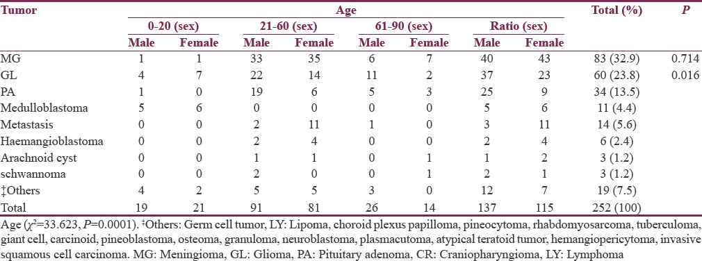
This study reviewed 252 patients out of 612 neuroimaging diagnosed brain tumors. Mean age was 42.8 years and male-to-female ratio was 1.2:1.0. Annual frequency increased from 11 in 2006 to 55 in 2015. Metastatic brain tumors accounted for 5.6%, and infratentorial tumors represented 16.3%. Frequency of the common primary tumors were meningioma (32.9%), glioma (23.8%), pituitary adenomas (13.5%), and craniopharyngioma (7.5%) (P = 0.001). Vestibular schwannoma accounted for 1.2%. Meningioma did not have gender difference (P = 0.714). Medulloblastoma, glioma, and craniopharyngioma were the most common pediatric tumors. About 8.7% presented unconscious (P < 0.001). There was no significant difference between radiology and histology diagnosis (P = 0.932).
Conclusion:
Meningioma is the most frequent tumor with increasing male incidence, but the frequency of glioma is increasing. Metastasis, acoustic schwannoma, lymphoma, and germ cell tumors seem to be uncommon. Late presentation is the rule.
Keywords
Epidemiology
geographical-neurosurgery
intracranial-tumors
INTRODUCTION
There is increasing evidence that suggests geographical differences in the incidence and distribution of intracranial tumors (ICTs), just like other neurosurgical disorders worldwide.[12] The previous reports have drawn attention to the relatively lower incidences of some types of brain tumors in some parts of Africa when compared to other parts of the world.[3] Earlier publications from Nigeria reported that brain metastasis and astrocytoma were the most common brain tumors among the adult and pediatric age groups, respectively.[45] More recent reports from sub-Saharan Africa suggest an increasing incidence of meningioma among the brain tumors in the past two decades.[367] These findings are strikingly different from the Caucasians, where gliomas, especially glioblastoma predominate. It has been argued that the results from sub-Saharan Africa concerning the epidemiology of ICTs may have been influenced by factors such as shorter life expectancy, poor hospital attendance habit, sociocultural factors that may delay patient decision to seek expert care, dearth of expertise, and relevant facilities needed for proper diagnosis of brain tumors.[1] These factors may give a picture of an apparently low incidence of ICTs.
However, these circumstances have been changing in the past decade with improvements in medical awareness. Expertise and neuroimaging facilities have also become available in many centers in Nigeria. The average socioeconomic conditions and life styles are also changing. One expects, therefore, that these paradigm shifts would help to define the true frequency of ICTs managed in Nigeria. It is also expected that the availability of neuroimaging would increase the proportion of asymptomatic tumors being diagnosed and managed.
The major aim of this study is to analyze the current distribution of histologically confirmed brain tumors managed in Enugu, Nigeria, over a decade.
METHODS
This is a retrospective analysis of prospectively recorded data of patients who were managed for ICTs at Memfys Hospital for Neurosurgery (MHN), Enugu, Nigeria. MHN was the major referral center for neuroimaging investigations and neurosurgery service in South-East and South-South zones of Nigeria during the greater part of this study. The study period spanned from January 2006 to December 2015. MHN had an 8-slice ceretom computed tomography (CT) machine throughout the duration of this study, a 64-slice Philips CT machine in the past 2 years, and 0.35T BASDA magnetic resonance imaging machine in the last 6 years of the study. Only patients that had surgery and conclusive postoperative histology report of an ICT were enlisted in the study. Nontumor space occupying lesions were excluded from the study. Relevant data were obtained from the medical records, radiology, and operation registers as well as histopathology reports. The age, sex, relative frequency of the tumors, patient presenting complaints, pre-operative imaging diagnosis, and anatomical location of tumor were analyzed. The date of diagnosis was based on the date of the preoperative imaging investigation. The tumors were classified based on the World Health Organization classification. Analysis was performed using descriptive and inferential statistics using tables, graphs, and Chi-square test with the aid of Statistical Package for Social Sciences (version 17 SPSS Inc., Chicago, IL, USA) software.
RESULTS
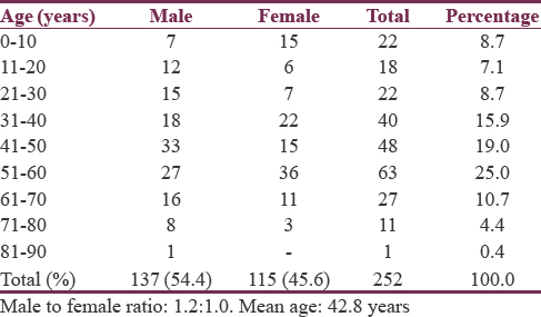
This study reviewed 252 patients managed with histology confirmed ICTs out of the 612 ICTs diagnosed from neuroimaging investigations during the study period. Mean age was 42.8 years. Male-to-female ratio was 1.2:1.0. Peak age range was the fifth (19.0%) to sixth (25.0%) decade with marked drop after the seventh decade of life (4.8%). The peak age incidence was the fifth decade in males and sixth decade in females [Table 1]. The average annual case presentations rose from 11 in 2006 to 55 in 2015, with peak month of presentation being in August while December to January period had the least case presentation frequency annually [Figures 1 and 2].
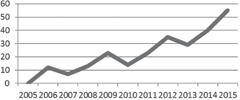
- Tumor frequency over the years
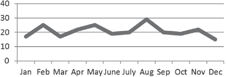
- Presentation pattern over the months
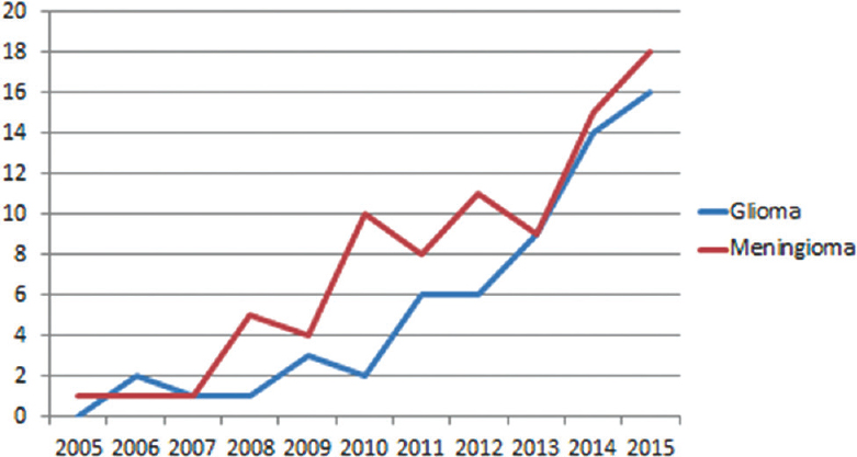
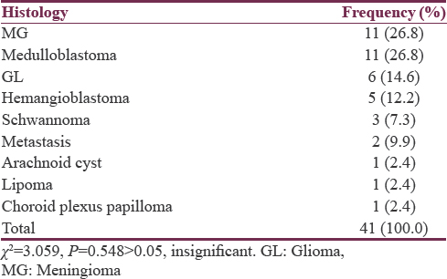
Primary ICTs accounted for 94.4% of the cases. Meningioma (32.9%), gliomas (23.8%), pituitary adenomas (13.5%), and craniopharyngioma (7.5%) were the most common tumors [Table 2]. Although meningioma is still the most common tumor, the relative proportion of glioma has been rising year on year in the past 4 years [Figure 3]. Medulloblastoma represented 4.4% of ICTs, hemangioblastoma 2.4%, arachnoid cyst 1.2%, and schwannoma 1.2% [Table 2]. In the pediatric age group, medulloblastoma (28.9%), glioma (23.6%), and craniopharyngioma (23.6%) were the most common tumors managed. There was male preponderance for pituitary adenoma (2.7:1) and glioma (1.6:1) at P = 0.016, while meningioma did not have significant gender preference (1:1.075) at P = 0.134 [Table 2]. Infratentorial tumors accounted for 16.3% of cases, and their order of frequency was meningioma (26.8%), medulloblastoma (26.8%), glioma (14.6%), and hemangioblastoma (12.2%). Ten percent of the posterior fossa tumors were metastasis [Table 3].
- Proportion of meningioma and glioma
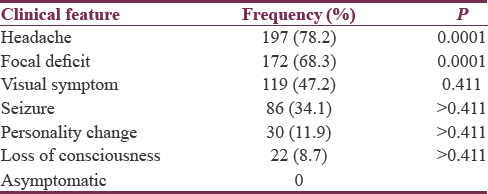
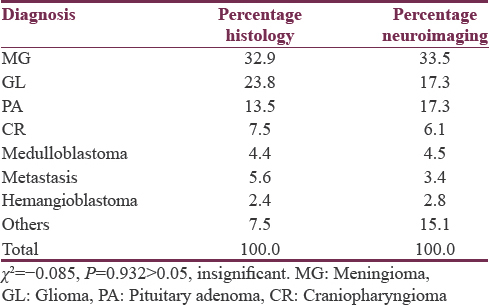
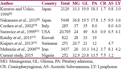
The presentation was varied [Table 4]. Headaches (78.2%), focal neurological deficit (68.3%), visual complaints (47.2%), and seizure (34.1%) were the most common presenting complaints, whereas 8.7% of the patients presented after alteration in the level of consciousness. There was no significant difference between radiology and histological diagnosis at P = 0.932 [Table 5]. Table 6 compares the relative distribution of different ICTs reported in different parts of the world with the current study.
DISCUSSION
This study has given an insight into the current epidemiology of ICTs in South-East Nigeria. The pattern of patient presentation over the years reveals a progressive increase in the number of patients presenting for surgery with histology confirmed brain tumors. This may be connected with gradual improvement in the level of awareness of brain tumors in the study environment. The increase in frequency of histology confirmed ICTs may also be an indication that the epidemiology of these tumors is still evolving. Although meningioma was the most common tumor managed, the proportion of gliomas managed yearly over the past 4 years was almost equal to meningioma. This would suggest that glioma in this environment is not as rare as previously believed. The recent rise in the relative frequency of gliomas may partly be due to improved access to neurosurgery and radiology services.
On the overall scale, however, the higher proportion of meningioma in this study is also similar to the pattern of brain tumors seen in Nigeria and some other parts of Africa where published reports in the past two decades show a trend toward higher incidences of meningioma relative to gliomas.[671516] Although factors such as life expectancy should be considered, the relative low glioma incidence from this study compared with the Western countries where glioblastoma prevalence is very high, especially among the elderly might suggest racial differences.[11] This study observed a very low incidence of tumors among the elderly, an age range where the incidence of gliomas should be high. Although life expectancy at birth among Nigerians has changed from about 45 years in the 1980s to the present 52 years, the relatively low incidence of gliomas may have a strong relationship with this rather low life expectancy. Nakamura et al.[9] observed a recent trend of increasing glioma incidence in Japan attributed to improved life expectancy and medicare services. Furthermore, this study observed that as high as 34% of the patients present with seizure as a major symptom. Generally, seizures are underinvestigated in Nigeria,[1718] and it may be possible that there are many cases of low-grade and high-grade gliomas being missed out as a result of inadequate seizure investigation.
Vestibular schwannoma and lymphoma are uncommon in this environment unlike other parts of the world.[8910111419] This is surprising, especially for lymphoma, considering the high prevalence of HIV/AIDS in this population and Burkitt lymphoma in the pediatric age group. The low incidence of both tumor types may be a true reflection of geographical differences and requires more detailed study. Although the role of neurosurgery in the treatment of lymphoma has reduced over the years due to effective alternative treatment options, it would have been expected that if the incidence of lymphoma was truly high, more cases should be picked up from neuroimaging studies and at least be confirmed from biopsy results for tumors. Similarly, germ cell tumors are rare in this report as is the trend in the Middle East, United States, and Italy but reported to be high among the Japanese population.[101420] Pediatric patients from sub-Saharan Africa appear to have a strong predisposition to medulloblastoma as also shown in this study. This trend is quite different in reports from North Africa and the Middle East.[1419]
A significant proportion of the patients presented at a relatively young age in this study. Environmental pollutions from oil and gas resources and improper disposal of industrial waste products are common in Nigeria, and these may increase the risks of nongerm line mutations in favor of a more aggressive tumor growth.[1918] Further studies need to be carried out to identify some peculiar genetic and nongenetic predisposing factors in this environment that may account for relatively younger age at patient presentation. It may be useful to also look at the biological behavior of the tumors managed to assess if the individuals in this locality present with more aggressive diseases, especially the malignant tumors.
The overall higher incidence of tumor in males is comparable to other studies worldwide although some studies from South America revealed a higher female tumor rate.[13] A peculiar finding in this study was the absence of gender difference in the distribution of meningioma and the clear male dominance of pituitary adenomas. The reason for this relative high male incidence is not clear, and a true geographical difference must be considered. In the pediatric age group, medulloblastoma was the most common pediatric tumor followed by craniopharyngioma. Craniopharyngioma was also the most common supratentorial pediatric tumor. Another study in this region reported similar pattern among the pediatric age group.[7] However, in Morocco and Italy, medulloblastoma followed by astrocytoma is the most common pediatric ICTs. In the Middle East, astrocytoma is the most common followed by medulloblastoma, whereas in Japan, astrocytoma is the most common pediatric ICT followed by germ cell tumors and then craniopharyngioma.[8101419] In the North America, craniopharyngioma was also more common in African-Americans than Caucasians.[202122]
None of the cases in this study was diagnosed incidentally. Headache and visual symptoms were very common.[20] Headache is a sensitive symptom which should not always be attributed to benign causes. Most other more specific localizing neurological deficit would manifest only after brain damage has occurred from advancing disease. Improved medical and social orientation in this direction may shorten the time between onset of symptom and presentation for expert care.
In developed countries, clinically silent tumors are being diagnosed with increasing frequency. Compared with the current study, this may be a reflection of the disparity in the level of organization of Medicare and health-seeking behavior of the population. It is expected that the current effort to increase the coverage of national health insurance scheme may improve the situation.
The major limitation of this study is that it is hospital based. In addition, this study reviewed only patients with histologically confirmed ICT out of the 612 ICT diagnosed from neuroimaging investigations during the study period and does not reflect the overall tumors operated on. From Table 5, it can be appreciated that there was a strong correlation between the histology and radiology diagnosis and a wider study based on radiology alone may provide more reliable epidemiology and may be indicated.
ICTs are still understudied in Nigeria and many African countries. A properly organized and funded community-based study or multi-center collaboration will definitely throw more light on the true incidence of ICTs. Despite this limitation, this study will help to strengthen the local literature on the epidemiology of ICTs in Nigeria. Hopefully, the newly introduced active cancer registry will also help with proper record keeping of tumors generally.
CONCLUSION
Significant differences in the epidemiology of ICTs are reported in this series from Enugu, Nigeria. Very few metastatic tumors were recorded. Medulloblastoma and craniopharyngioma are the predominant pediatric ICTs. Glioma and pituitary adenoma have marked male predominance while meningioma did not have a gender difference. Meningioma remains the most frequent tumor both from radiology- and histology-based analysis and shows increasing male incidence in this environment. Glioma and pituitary adenoma are also common, and the rate of glioma diagnosis is almost equivalent to meningioma in the past 4 years. However, vestibular schwannoma, lymphoma, and germ cell tumors are relatively uncommon in this environment. The geographical differences reported here may be due to genetic or environmental factors, a reflection of disparity in quality, and level of access to medicare and the disease reporting system in different parts of the world.
Financial support and sponsorship
Nil.
Conflicts of interest
There are no conflicts of interest.
Acknowledgment
The authors would like to thank Mr. Ade for statistical analysis.
REFERENCES
- Brain tumours: Incidence, survival, and aetiology. J Neurol Neurosurg Psychiatry. 2004;75(Suppl 2):ii12-7.
- [Google Scholar]
- Current epidemiological trends and surveillance issues in brain tumors. Expert Rev Anticancer Ther. 2001;1:395-401.
- [Google Scholar]
- Diagnosis and management of brain tumours at Jos University Teaching Hospital, Nigeria. East Afr Med J. 2001;78:148-51.
- [Google Scholar]
- Management of intracranial meningiomas in Enugu, Nigeria. Surg Neurol Int. 2012;3:110.
- [Google Scholar]
- Symptomatic primary intracranial neoplasms in Nigeria, West Africa. J Neurol Sci Turk. 2007;24:212-8.
- [Google Scholar]
- Epidemiological study of primary intracranial tumors in childhood. A population-based survey in Kumamoto Prefecture, Japan. Pediatr Neurosurg. 1996;25:240-6.
- [Google Scholar]
- Epidemiological study of primary intracranial tumors: A regional survey in Kumamoto prefecture in Southern Japan-20-year study. Int J Clin Oncol. 2011;16:314-21.
- [Google Scholar]
- Epidemiology of primary intracranial tumours in NW Italy, a population based study: Stable incidence in the last two decades. J Neurol. 2002;249:281-4.
- [Google Scholar]
- Descriptive epidemiology of primary brain and CNS tumors: Results from the Central Brain Tumor Registry of the United States, 1990-1994. Neuro Oncol. 1999;1:14-25.
- [Google Scholar]
- Incidence and treatment of central nervous system tumors in Suriname. World Neurosurg. 2013;80:e79-83.
- [Google Scholar]
- Epidemiology of primary intracranial tumors in Iran, 1978-2003. Asian Pac J Cancer Prev. 2006;7:283-8.
- [Google Scholar]
- Factors influencing visual and clinical outcome in Nigerian patients with cranial meningioma. J Clin Neurosci. 2006;13:649-54.
- [Google Scholar]
- Neuroimaging findings in pediatric patients with seizure from an institution in Enugu. Niger J Clin Pract. 2016;19:121-7.
- [Google Scholar]
- Epilepsy in primary intracranial tumors in a neurosurgical hospital in Enugu, South-East Nigeria. Niger J Clin Pract. 2015;18:681-6.
- [Google Scholar]
- Epidemiologic profile of pediatric brain tumors in Morocco. Childs Nerv Syst. 2010;26:1021-7.
- [Google Scholar]
- The presenting features of brain tumours: A review of 200 cases. Arch Dis Child. 2006;91:502-6.
- [Google Scholar]
- 2001 CBTRUS Statistical Report: Primary Brain Tumors in the United States, 1992-97. Available from: http://www.cbtrus.org/2001/2001stats_report.htm
- Canadian Cancer Society's Steering Committee for Canadian Cancer Statistics. Canadian cancer statistics at a glance: Cancer in children. CMAJ. 2009;180:422-4.
- [Google Scholar]


