Translate this page into:
Diagonal Earlobe Crease Revealing Intracranial Atherosclerosis
Address for correspondence: Dr. Oscar H. Del Brutto, Air Center 3542, PO Box 522970, Miami, FL 33152-2970, USA. E-mail: oscardelbrutto@hotmail.com
This is an open access journal, and articles are distributed under the terms of the Creative Commons Attribution-NonCommercial-ShareAlike 4.0 License, which allows others to remix, tweak, and build upon the work non-commercially, as long as appropriate credit is given and the new creations are licensed under the identical terms.
This article was originally published by Medknow Publications & Media Pvt Ltd and was migrated to Scientific Scholar after the change of Publisher.
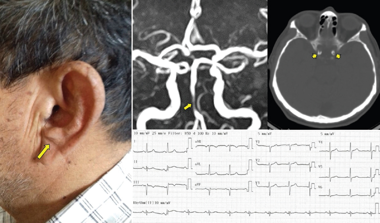
An asymptomatic 76-year-old man with arterial hypertension and an old myocardial infarction was evaluated after providing signed informed consent for being enrolled in the Atahualpa Project, a population-based cohort study designed to reduce the increasing burden of noncommunicable cardiovascular and neurological diseases in rural Ecuador.[1] Physical examination was remarkable for the presence of a diagonal crease in the left earlobe (known as Frank's sign) [Figure 1, left], which has been associated with increased risk of atherosclerotic disease.[2] A 12-lead electrocardiogram confirmed the presence of an inferior myocardial infarction [Figure 1, right lower panel]. Time-of-flight magnetic resonance angiography showed severe segmental stenosis of the basilar artery, and high-resolution computed tomography with bone window settings revealed the presence of high calcium content in both carotid siphons (a recognized surrogate of intracranial atherosclerosis) [Figure 1, right upper panel].

- Photograph of the patient showing a diagonal earlobe crease (large arrow), magnetic resonance angiography showing severe segmental stenosis of the basilar artery (short arrow), computed tomography showing severe calcification of both carotid siphons (arrowhead), and electrocardiogram showing an old inferior myocardial infarction
Diagnosis of most neurological diseases requires the use of sophisticated technology, which is not available in rural areas. The Frank's sign might be used as a simple tool to identify candidates for the practice of neuroimaging studies in research studies conducted in remote rural settings. This will help identify apparently healthy individuals at risk for developing catastrophic diseases before they occur.
Declaration of patient consent
The authors certify that they have obtained all appropriate patient consent forms. In the form the patient(s) has/have given his/her/their consent for his/her/their images and other clinical information to be reported in the journal. The patients understand that their names and initials will not be published and due efforts will be made to conceal their identity, but anonymity cannot be guaranteed.
Financial support and sponsorship
This study was partly supported by an unrestricted grant from Universidad Espíritu Santo, Ecuador, Guayaquil, Ecuador.
Conflicts of interest
There are no conflicts of interest.
REFERENCES
- Door-to-door survey of cardiovascular health, stroke, and ischemic heart disease in rural coastal ecuador – the atahualpa project: Methodology and operational definitions. Int J Stroke. 2014;9:367-71.
- [Google Scholar]
- Anterior tragal crease is associated with atherosclerosis: A Study evaluating carotid artery intima-media thickness. Angiology. 2017;68:683-7.
- [Google Scholar]


