Translate this page into:
Acute inter-hemispheric subdural hematoma in a kabaddi player: A comment
Address for correspondence: Dr. M A Hashmi, MRI-Section, EKO CT and MRI Scan Centre at Medical College And Hospitals Campus, 88, College St, Kolkata - 700 073, India. E-mail: ahashmidrrad@yahoo.co.ine
This is an open-access article distributed under the terms of the Creative Commons Attribution-Noncommercial-Share Alike 3.0 Unported, which permits unrestricted use, distribution, and reproduction in any medium, provided the original work is properly cited.
This article was originally published by Medknow Publications and was migrated to Scientific Scholar after the change of Publisher.
Sir,
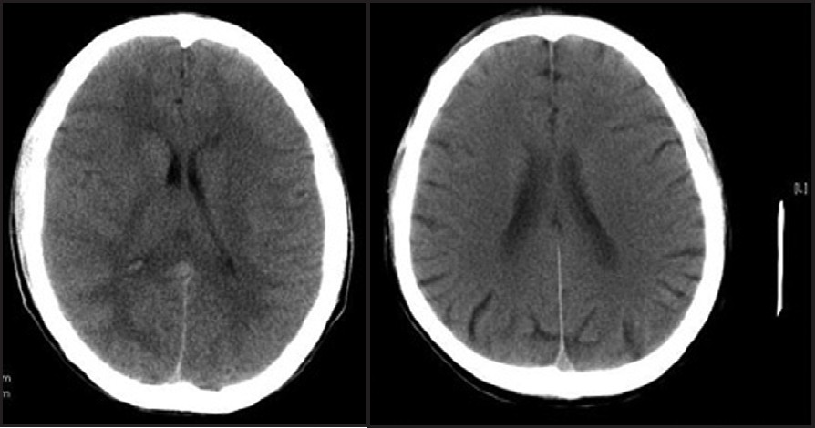
I had read with great interest the recent publication on acute inter-hemispheric subdural hematoma in a kabaddi player.[1] I have some comment on this work. First, the image as shown in Figure 1 in the original article has been described as inter-hemispheric subdural hematoma. According to me differential can be partial volume of straight and sagittal sinuses, commonly seen as hyperdense in plain CT scan imaging. Figure 1 shows plain CT scan of the brain of two different patients; partial volume of the sinuses can be seen there. Second, the posterior portion of the image in figure in the original article appears as inverted Y appearance when it is following the course of transverse sinuses. Subdural hematoma will never or rarely have inverted Y-shaped most posterior end. Whether an MRI with angio- and veno-sequence was done to rule out above condition is not clear, and no remark has been done on follow-up imaging whether it was done or not.

- Plain CT scan of the brain of two different patients shows partial volume of sagittal and straight sinuses as hyperdensity in the posterior interhemispheric fissure.
Reference
- Acute inter-hemispheric subdural haematoma in a kabaddi player. J Neurosci Rural Pract. 2010;1:122-3.
- [Google Scholar]


