
Translate this page into:
Solitary fibrous tumor of the thoracic spine
Address for correspondence: Dr. Rafael Cincu, Department of Neurosurgery, Valencia, Spain. E-mail: rafael.cincu@gmail.com
This is an open-access article distributed under the terms of the Creative Commons Attribution-Noncommercial-Share Alike 3.0 Unported, which permits unrestricted use, distribution, and reproduction in any medium, provided the original work is properly cited.
This article was originally published by Medknow Publications and was migrated to Scientific Scholar after the change of Publisher.
Introduction
Solitary fibrous tumors (SFT) are uncommon mesenchymal tumors that involve the pleural cavity and numerous extrathoracic sites, including prostate, kidney, thyroid, and rarely the spinal cord.[1–6] A 58-year-old white male was admitted with complaints of progressive weakness and sensory disturbance of the lower extremities of 2-year duration. There was no history of fever or trauma. His bowel and bladder functions were normal. His general and systemic examination was normal. Higher mental functions, cranial nerves, and motor and sensory functions in the upper limbs were normal. Further neurologic examination revealed decreased pain, light touch, and vibration sensations below the D4 level, muscle weakness of grade 4/5 of all muscle groups in lower limbs. The deep tendon reflexes were exaggerated in both the lower limbs, and the bilateral planters were extensor. His complete blood count and relevant clinical chemistry was normal. Magnetic resonance imaging of the thoracic spine revealed a dorsally placed well-defined mass extending from D3 to D5 level, compressing the spinal cord. The mass was mildly hypointense on the T1-weighted images and hyperintense on T2-weighted images [Figure 1]. The patient underwent D3–D5 laminectomy and tumor extirpation. There was intradural milky-white, noncapsulated tumor dorsal to the spinal cord with a definite plane of cleavage between the tumor and the surrounding cord tissue. The tumor could be removed completely. Microscopic examination of the specimen revealed spindle cell proliferation. On immunohistochemical study [Figure 2], the tumor cells were positive for vimentin, but negative for bcl2— the findings compatible with SFT.[15–8] The patient has been doing well at follow-up. SFT arising in the spinal cord needs to be differentiated from schwannoma, meningioma, and hemangioblastoma.[1–5] Although imaging modalities can provide a preliminary indication, the diagnosis of SFT is usually made on histology.[1269] A careful morphologic approach and the judicious use of immunohistochemistry may assist in distinguishing them among these conditions, although some irreducible difficulties may be faced by academics of taxonomy.[1–5] SFTs are usually indolent neoplasms and complete surgical resection of all involved tissue is recommended.[1–3] In view of the indolent nature of the SFTs, radiotherapy or chemotherapy for remnant disease is not recommended.[34]
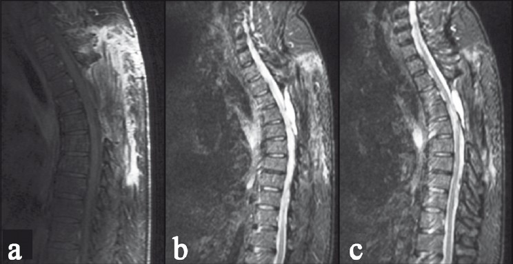
- Magnetic resonance imaging sagittal T2W images showing a hyperintense mass, placed dorsal to the cord extending from D3 to D5 level, with cord compression
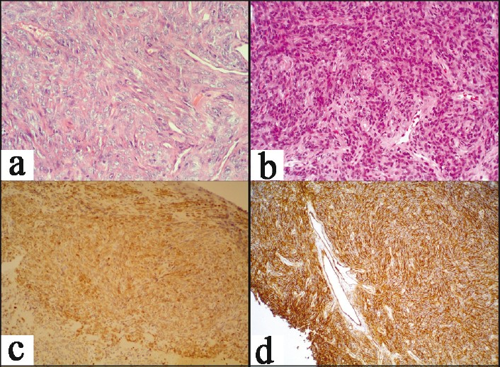
- Photomicrographs showing (a) spindle cell proliferation in interfascicular pattern with thin slit-like blood vessel, H and E ×20; (b) photomicrograph showing the hyperchromatic spindle cells with mild pleomorphism and infrequent mitoses, H and E ×40; (c) immunohistochemistry for bcl2-negative staining pattern; and (d) immunohistochemistry showing vimentin positivity among the tumor cells
Source of Support: Nil,
Conflict of Interest: None declared.
References
- [Polyostotic fibrous dysplasia of the thoracic spine. A case study and review of the literature] Neurochirurgie. 2009;55:595-9.
- [Google Scholar]
- A rare case of malignant solitary fibrous tumor of the spinal cord. Spine (Phila Pa 1976). 2008;33:E397-9.
- [Google Scholar]
- Spinal solitary fibrous tumors: A series of four patients: Case report. Neurosurgery. 2005;57:E195. discussion E195
- [Google Scholar]
- Solitary fibrous tumor of the spinal cord: Case report and review of the literature. Neurosurgery. 2004;55:433.
- [Google Scholar]
- Solitary fibrous tumor of the thoracic spinal cord. Neurol Med Chir (Tokyo). 2005;45:371-4.
- [Google Scholar]
- Solitary fibrous tumor of the spinal cord: A clinicopathologic study of two cases. Ann Diagn Pathol. 2004;8:268-75.
- [Google Scholar]
- Solitary fibrous tumors in the central nervous system.A clinicopathologic review of 18 cases and comparison to meningeal hemangiopericytomas. Arch Pathol Lab Med. 2003;127:432-9.
- [Google Scholar]
- A dumbbell-shaped solitary fibrous tumor of the cervical spinal cord. Yonsei Med J. 2008;49:167-70.
- [Google Scholar]


