
Translate this page into:
Inverted V or rabbit ear sign in cerebellum
Address for correspondence: Dr. Venkatesan Prasanna Eswaradass, 3, Vijayarahavachari Road, Gandhi Road, Salem - 636 007, Tamil Nadu, India. E-mail: eprasanna2k1@gmail.com
This is an open-access article distributed under the terms of the Creative Commons Attribution-Noncommercial-Share Alike 3.0 Unported, which permits unrestricted use, distribution, and reproduction in any medium, provided the original work is properly cited.
This article was originally published by Medknow Publications & Media Pvt Ltd and was migrated to Scientific Scholar after the change of Publisher.
We report a 55-year-old female who presented to us with history of sudden onset of vertigo, vomiting and ataxia for 2 days duration. The patient is a known hypertensive on regular treatment for last 7 years. She was also a recently detected diabetic on oral hypoglycemic therapy. There was no past history of myocardial infarction, transient ischemic attack (TIA) or stroke. On examination, she had bilateral gaze evoked nystagmus, bilateral finger nose incoordination and gait ataxia. There was no long tract signs to suggest involvement of brainstem. All peripheral pulses were felt and regular in rhythm. Clinically, cardiovascular system was normal. MR imaging of brain revealed acute infarts in inferior cerebellum in the bilateral medial PICA (posterior inferior cerebellar artery) territories [Figure 1]. Magnetic resonance angiogram (MRA) did not reveal any abnormality. Digital subtraction angiography (DSA) showed bilateral occlusion of PICA. Vertebral arteries were normal [Figure 2]. She made good recovery without any complications and was discharged with minimal gait ataxia after 10 days.
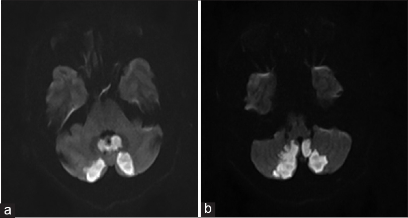
- (a and b) MRI DWI shows bilateral inverted V shaped infarct in PICA territory
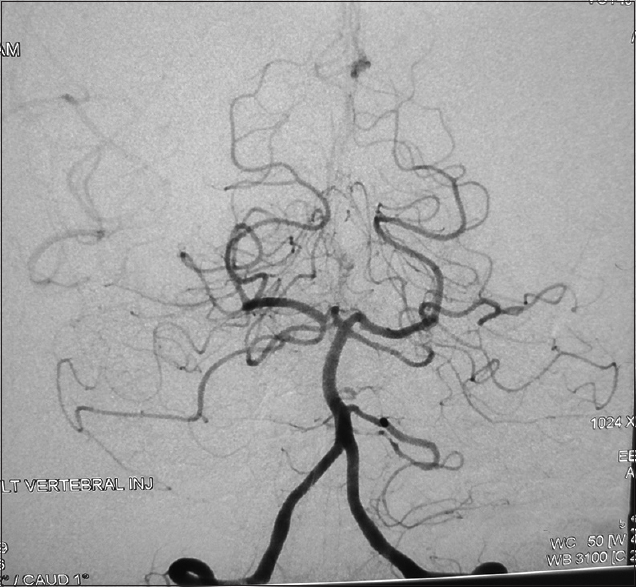
- DSA shows bilateral PICA and right AICA occlusion
Inverted “V” or rabbit ear sign is a well-known neuroradiological sign seen in subacute combined degeneration (SACD) of spinal cord due to vitamin B12 deficiency. We encountered a rare case of inverted “V” or rabbit ear sign in cerebellum. Cerebellum is supplied by PICA, anterior inferior cerebellar artery and superior cerebellar artery. PICA is the largest branch of vertebral artery and divides into medial and lateral branch and supplies most of inferior surface of cerebellum and medulla. Simultaneous bilateral cerebellar infarctions in PICA territory, without brain stem involvement, are rare. Bilateral PICA infarcts may be due to anatomical variant. It is worth emphasizing that of all the major arteries of the human brain, the PICAs have the most varied anatomy.[1] It is possible that both PICAs originate from an occluded basilar artery or both medial branches arise from the single PICA. Rarely double embolic stroke affecting both medial PICA can cause this type of infarction.[2]
In our case, DSA showed non-opacification of bilateral PICA with normal vertebral arteries and did not reveal any anatomical variant. The hypothesis made by Kang et al. may fit this case. On one side, PICA could be a hypoplastic one, resulting in non-opacification. On the other side, dominant PICA (supplying bilateral medial PICA territories) could have been occluded, resulting in bilateral medial PICA territory infarcts.[3] As the bilateral infarcts are simultaneous and bilaterally symmetrical, this hypothesis can be considered apart from an embolic etiology.
Neurologist must be aware of rare bilateral simultaneous medial PICA infarcts, as they may initially present only with ataxia or vertigo and may worsen subsequently due to edema of the infarct. The resultant pressure on the brainstem and obstruction of CSF flow can cause progressive coma.[4] Bilateral PICA infarction in cerebellum can mimic inverted “V” sign of SACD. It is a rare syndrome and probably occurs due to anatomical variant or due to embolic stroke.
Source of Support: Nil.
Conflict of Interest: None declared.
References
- La vascularisationarterielle du cervelet. In: Lazorthes G, ed. Vascularisationet Circulation Cerebrales. Paris, France: Masson; 1961. p. :65-75.
- [Google Scholar]
- Bilateral simultaneous cerebellar infarction in the medial branches of the posterior inferior cerebellar artery territories. Ital J Neurol Sci. 1996;17:433-6.
- [Google Scholar]
- Acute bilateral cerebellar infarcts in the territory of posterior inferior cerebellar artery. Neurology. 2000;55:582-4.
- [Google Scholar]
- Acute bilateral inferior cerebellar infarction in a patient with neurosyphilis. Arch Neurol. 2004;61:953-6.
- [Google Scholar]


