Translate this page into:
Facial Diplegia as Initial Manifestation of Acute, Myelomonocytic Leukemia with Isolated Trisomy 47, XY,+11[14]/46, XY[6]
Address for correspondence: Dr. Josef Finsterer, Postfach 20, 1180 Vienna, Austria. E-mail: fifigs1@yahoo.de
This is an open access article distributed under the terms of the Creative Commons Attribution-NonCommercial-ShareAlike 3.0 License, which allows others to remix, tweak, and build upon the work non-commercially, as long as the author is credited and the new creations are licensed under the identical terms.
This article was originally published by Medknow Publications & Media Pvt Ltd and was migrated to Scientific Scholar after the change of Publisher.
Abstract
Bilateral peripheral facial palsy (facial diplegia) has been repeatedly reported as a neurologic manifestation of acute myeloid leukemia but has not been reported as the initial clinical manifestation of myelomonocytic leukemia. A 71-year-old male developed left-sided peripheral facial palsy being interpreted and treated as Bell's palsy. C-reactive protein (CRP) and leukocyte count 4 days later were 2.5 mg/l and 16 G/l, respectively. Steroids were ineffective. Seven days after onset, he developed right-sided peripheral facial palsy. Three days later, CRP and leukocyte count were 234.3 mg/l and 59.5 G/l, respectively. Cerebrospinal fluid investigations revealed pleocytosis (62/3) and elevated protein (54.9 mg/dl). Two days later, pleocytosis and leukocytosis were attributed to myelomonocytic leukemia. Leukemic meningeosis was treated with cytarabine and methotrexate intrathecally. In addition, cytarabine and idarubicin were applied intravenously. Under this regimen, facial diplegia gradually improved. Facial diplegia may be the initial clinical manifestation of myelomonocytic leukemia, facial diplegia obligatorily requires lumbar puncture, and unilateral peripheral facial palsy is not always Bell's palsy. Patients with alleged unilateral Bell's palsy and slightly elevated leukocytes require close follow-up and more extensive investigations than patients without abnormal blood tests.
Keywords
Bell's palsy
chemotherapy
facial palsy
leukemia
leukemic meningeosis
INTRODUCTION
Facial palsy is a common disease with a favorable outcome in the majority of the cases. Usually, it occurs unilaterally, but rarely bilateral facial palsy (facial diplegia) has been reported. The most common cause of unilateral facial palsy is Bell's palsy. The most common cause of facial diplegia is sarcoidosis, but there are a number of rare other causes of facial diplegia [Table 1]. One of the rare causes of facial diplegia is leukemia. Facial diplegia has been reported as a neurologic manifestation of acute myeloid leukemia (AML)[123] and of acute lymphoid (lymphoblastic) leukemia (lymphoma).[4567] Facial diplegia from leukemia may be due to mastoid infiltration,[1] due to leukemic meningitis,[8] or due to bilateral focal lesion of the facial nucleus. Depending on the location of the infiltration with leukemic cells, the cerebrospinal fluid (CSF) may be normal or may show leukemic cells as well. Although facial diplegia has been reported as initial manifestation of acute T-cell leukemia,[8] it has not been reported as initial manifestation of acute myelomonocytic leukemia.

CASE REPORT
A 71-year-old Caucasian male presented at the ambulatory ward with left-sided peripheral facial palsy with incomplete lid closure after a bronchial infection without taking antibiotics 4 days before. Bell's palsy was diagnosed, and treatment with steroids (25 mg prednisolone over 10 days) and physiotherapy was initiated. Three days later, he also developed a right-sided peripheral facial palsy of the same degree. Another 3 days later (6 days after the first visit), he again attended the ambulatory ward (hospital day [hd] 1). Blood tests showed increased C-reactive-protein and white blood cell (WBC) counts [Table 2]. Cerebral magnetic resonance imaging (MRI) revealed mild diffuse atrophy and multiple, gliotic spots in a bilateral frontotemporal distribution. CSF investigations showed mild pleocytosis and elevated protein [Table 2]. Due to a history of recurrent tick bites over the last years, he additionally received ceftriaxone (2 g/d) intravenously during 7 days and acyclovir (1500 mg/d) intravenously during 7 days. His previous history further revealed cervical disc extraction in 1981 complicated by recurrent abscess formation and antibiotic treatment until 2010, lumbar disc extraction in 2006, prostatectomy in 2009, knee endoprosthesis in 1/2014, hyperuricemia, and phimosis. In addition to facial diplegia, neurologic examination showed bilateral hypoacusis, mild weakness for hip flexion on the right side, and generally reduced tendon reflexes.

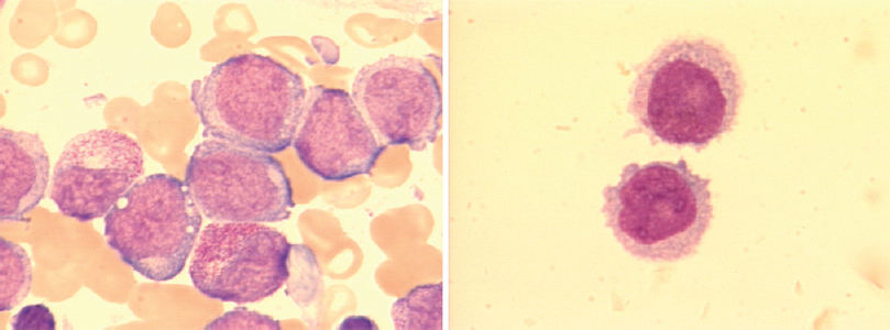
Cervical MRI on hd 2 revealed paradox kyphosis, anterolisthesis C3/4 with consecutive vertebrostenosis and stenosis of the neuroforamina, partial block vertebrae C4–6 with resected spinous processes, discrete myelopathy, hydromyelia C3–7, an enhancing, intramedullary mass lesion C5, and bone marrow edema C7/Th1. Computed tomography scans of the thorax and the abdomen revealed extensive pneumonia, mediastinal and hilar lymphadenopathy, mild pericardial effusion, and hepatosplenomegaly. Differential WBC count showed a population of approximately 80% immature monocytic cells. Therefore, the patient was transferred to the hematological unit on hd 3 where myelomonocytic leukemia was diagnosed upon bone marrow analysis on hd 4 [Figure 1]. Further CSF analysis showed 69/3 cells, expressing various immature myeloid antigens (HLA-DR+, CD13+, CD117+, MPO+, and LZ+) confirming the suspected leukemic meningeosis. Cytogenetic investigations revealed isolated trisomy 11 in 14 of 20 analyzed metaphases (karyotype: 47, XY,+11[14]/46, XY[6]). Since hd 7, the patient developed mild quadriparesis starting on the left upper limb (M3). Leukemic meningitis was treated with cytarabine (40 mg), methotrexate (15 mg), and dexamethasone (4 mg) intrathecally on hd 4 and hd 17. Systemic antileukemic therapy consisted of cytarabine (100 mg/m2/day for 7 days) and idarubicin (12 mg/m2 on 2 days) according to the 2 + 7 scheme between hd 4 and hd 10. This treatment led to the improvement of facial diplegia. Although the leukocyte count in the CSF continuously declined [Table 2], quadriparesis did not improve before hd 13. On hd 37, bone marrow aspiration demonstrated partial remission.

- Leukemic blast cells of the patient in bone marrow (left panel) and spinal fluid (right panel)
DISCUSSION
The presented case is interesting for several aspects. First, it is a rare case of facial diplegia. In general, facial diplegia may be caused by a number of different conditions including infections, vascular problems, and immunological abnormalities [Table 1], of which sarcoidosis is the most frequent.[11] Second, facial diplegia was due to meningeal spread of myelomonocytic leukemia. Although facial diplegia has been repeatedly reported as a manifestation of AML or lymphoid leukemia,[1234567] only one case has been reported so far, in which this neurologic presentation occurred as a primary manifestation before the diagnosis of acute myelomonocytic leukemia.[2] Only unilateral facial palsy has been reported in association with myelomoncytic leukemia.[12] Only in a single case with T-cell leukemia was facial diplegia the initial manifestation of the disease.[7] Compared to lymphoid leukemia, myelomonocytic leukemia carries an increased risk of extramedullary involvement including spreading to the central nervous system (CNS). Third, the patient carried an isolated trisomy of chromosome 11, which occurs in <1% of the cases with AML.[13] Extramedullary manifestations have been described in these patients but never leukemic meningeosis. Disregarding the extent of the spreading, the prognosis of these patients is poor.[13]
The cause of quadriparesis remains unclear, but limb weakness was attributed either to the spinal lesion at C5 being interpreted as a leukemic focus or due to infiltration of the spinal roots or the plexus bilaterally by monoblasts. Infiltration of the plexus by leukemic cells has been previously reported in monoblastic leukemia.[14] Arguments for a C5 lesion are that it was not found on previous imaging studies and that CNS infiltration has been previously reported in myelomonocytic leukemia.[15] An argument against the C5 lesion is that clinically not only C5-innervated muscles were affected. Arguments for radiculopathy or bilateral plexopathy are that it has been previously described as a manifestation of leukemia,[1617] that tendon reflexes were generally reduced, and that it would explain quadriparesis. An argument against plexopathy, however, is that sensory functions were intact.
The reason why monoparesis of the left upper limb did not immediately respond to chemotherapy remains unknown, but several speculations can be raised to explain drug resistance. First, the follow-up interval after initiation of treatment was too short to observe a beneficial effect. Second, upper limb weakness was not due to leukemia but due to other causes, such as spinal stroke, a paraneoplastic phenomenon, or due to radiculitis or plexopathy from infectious or noninfectious causes. Third, the dosage of chemotherapeutics reaching the intraspinal lesion was too low to exhibit a beneficial effect. In case monoparesis was due to a plexus lesion, it is conceivable that chemotherapeutics were not able to cross the blood-nerve barrier why monoblasts could have escaped from chemotherapeutic agents.[12]
This case shows that facial diplegia may be the initial clinical manifestation of myelomonocytic leukemia and that unilateral peripheral facial palsy is not always Bell's palsy. Patients with alleged unilateral Bell's palsy, refractory or progressive unilateral facial palsy, or facial diplegia require close follow-up and more extensive investigations including differential blood counts and lumbar and bone marrow punctures.
Financial support and sponsorship
Nil.
Conflicts of interest
There are no conflicts of interest.
REFERENCES
- Facial diplegia and mastoid infiltration as symptoms of relapsed acute myeloid leukemia. Eur Ann Otorhinolaryngol Head Neck Dis 2014 pii: S1879-7296(14) 00036-2
- [Google Scholar]
- Bilateral facial palsy and acute myeloid leukemia: An unusual association. Acta Med Port. 2012;25:250-3.
- [Google Scholar]
- Bilateral facial nerve paralysis-diagnosis and treatment. Zhonghua Er Bi Yan Hou Tou Jing Wai Ke Za Zhi. 2006;41:821-4.
- [Google Scholar]
- Precursor T cell acute lymphoblastic lymphoma presenting as bilateral facial nerve palsy. Chemotherapy. 2010;56:258-60.
- [Google Scholar]
- Bilateral facial nerve palsy as first indication of relapsing hairy cell leukemia after 36 years. Neurology. 2004;63:399-400.
- [Google Scholar]
- An alarming sign for serious diseases in children: Bilateral facial paralysis. Pediatr Neurol. 2002;27:312-3.
- [Google Scholar]
- Facial diplegia as the presenting manifestation of acute lymphoblastic leukemia. Mt Sinai J Med. 2001;68:406-9.
- [Google Scholar]
- Adult T-cell leukemia initially manifesting as facial diplegia. Am J Hematol. 1989;32:61-5.
- [Google Scholar]
- Bilateral seventh nerve palsy: Analysis of 43 cases and review of the literature. Neurology. 1994;44:1198-202.
- [Google Scholar]
- Clinical aspects of bilateral facial paralysis. Laryngol Rhinol Otol (Stuttg). 1987;66:477-9.
- [Google Scholar]
- Bilateral facial paralysis: Case presentation and discussion of differential diagnosis. J Gen Intern Med. 2006;21:C7-10.
- [Google Scholar]
- Juvenile myelomonocytic leukemia presenting with facial nerve paresis: A unique presentation. J Pediatr Hematol Oncol. 2007;29:770-3.
- [Google Scholar]
- Trisomy 11 as an isolated abnormality in acute myeloid leukemia is associated with unfavorable prognosis but not with an NPM1 or KIT mutation. Int J Clin Exp Pathol. 2011;4:371-7.
- [Google Scholar]
- Infiltrative peripheral neuropathy of acute monoblastic leukemia during hematologic remission. Rinsho Ketsueki. 1994;35:876-80.
- [Google Scholar]
- Central nervous system lesions due to juvenile myelomonocytic leukemia progressed in a boy undergoing first line chemotherapy. Int J Hematol. 2012;95:581-4.
- [Google Scholar]
- Brachial plexopathy due to myeloid sarcoma in a patient with acute myeloid leukemia after allogenic peripheral blood stem cell transplantation. Ann Rehabil Med. 2013;37:280-5.
- [Google Scholar]
- Late relapse of acute myeloblastic leukemia as myeloid sarcoma causing radiculopathy. Leuk Res. 2010;34:e348-50.
- [Google Scholar]


