Translate this page into:
Extensive heterotopic ossification in patient with tubercular meningitis
Address for correspondence: Dr. Ganesh Yadav, Department of Physical Medicine and Rehabilitation, King George Medical University, Chowk, Lucknow - 226 003, Uttar Pradesh, India. E-mail: ganeshyadav4u@yahoo.co.in
This is an open-access article distributed under the terms of the Creative Commons Attribution-Noncommercial-Share Alike 3.0 Unported, which permits unrestricted use, distribution, and reproduction in any medium, provided the original work is properly cited.
This article was originally published by Medknow Publications & Media Pvt Ltd and was migrated to Scientific Scholar after the change of Publisher.
Abstract
Tubercular meningitis is a severe form of central nervous system tuberculosis with high morbidity and mortality. Apart from neurological deficits, musculoskeletal involvement is also seen in very few cases in the form of heterotopic ossification around immobile joints. A 35-year-old male case of tubercular meningitis with left hemiparesis presented with multiple joint restriction of range of motion. On clinical examination, palpable firm masses around multiple joints with painful restriction of movements were seen. X-ray films of multiple joints revealed heterotopic ossification over left shoulder, hip and knee joint with bony ankylosis of left hip and soft tissue contractures. Very few reports have been published in the literature for association of heterotopic ossification with tubercular meningitis with such extensive joint involvement which compels us to report this clinical association of tubercular meningitis. This report is intended to create caution among physicians and other caregivers for this debilitating complication of tubercular meningitis and in face of high prevalence of tuberculosis and tubercular meningitis, employ methods to prevent and treat.
Keywords
Heterotopic ossification
heterotopic ossification in hemiparesis
neurogenic heterotopic ossification
rehabilitation in heterotopic ossification
tubercular meningitis
Background
Heterotopic ossification (HO) is the abnormal bone formation within skeletal soft tissues. The term myositis ossificans is not used anymore because primary muscle inflammation is not a necessary precursor and ossification does not always occur in muscle tissue. It frequently shows a predilection for fascia, tendons and other mesenchymal soft tissues. Thus, term HO has replaced myositis ossificans in the literature.
It most commonly occurs in patients with complete injuries at a cephalad level. Periarticular HO is seen in patients with traumatic brain injury, tetanus, Gullian-Barre syndrome, Poliomyelitis, prolonged mechanical ventilation, severe trauma, hip arthroplasty and burns.[123456]
Central nervous system tuberculosis (CNS TB) comprises about 10% of all TB cases.[7] Tubercular meningitis (TBM) is the most common form of CNS TB, a subacute disease with high morbidity and mortality if left untreated. In survivors of TBM, neurological sequelae are common which may include hydrocephalus, cranial nerve palsies, stroke and associated neurological deficits, seizures, mental retardation, spasticity and coma.[8] TBM predominantly occurs in developing countries. These numbers are likely to rise due to an increase in drug resistance, increased use of immunosuppressive therapies, and increase in incidence of HIV.
Case Report
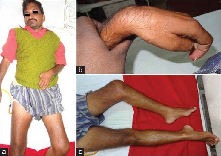
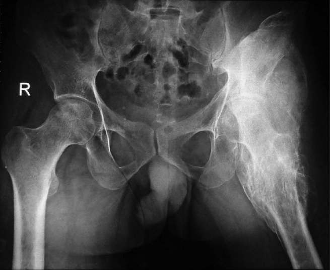
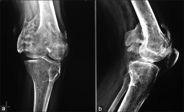
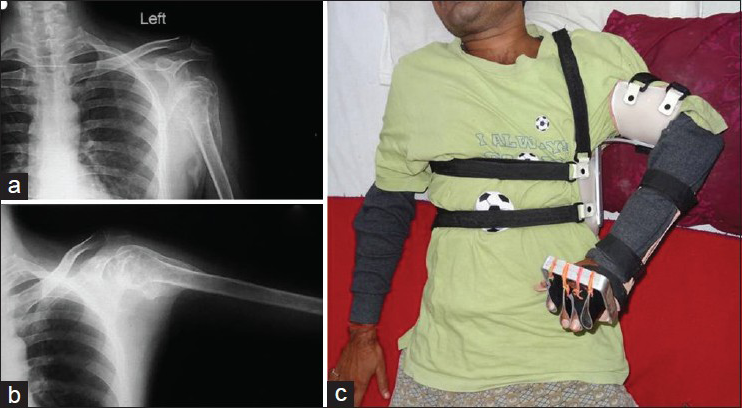
A 35-year-old right-handed male was admitted with diagnosis of TBM with hydrocephalus with left-sided hemiparesis. History revealed loss of consciousness 15 months back following complaint of low grade fever of long duration. He was managed by ventriculo-peritoneal shunt placement along with antitubercular therapy. The patient regained consciousness after 1 week but was unable to move his left upper and lower limb. Speech was slurred and sensations were intact bilaterally with no bladder or bowel involvement. Six months later, the patient developed progressive limitation of left hip and knee movement. A month later he suffered left shoulder pain during a physical therapy session, after which the patient and his caretakers stopped giving any therapeutic exercises due to severe pain on movements. Physical examination revealed contractures over his left shoulder, hip and knee [Figure 1] with ankylosis of left hip and knee [Figures 2 and 3]. Investigations revealed normal hemogram. Serum alkaline phosphatase was elevated. The radiographic examination revealed ectopic bone deposition around, left hip [Figure 2], left knee [Figure 3]and left shoulder [Figure 4]. MRI of brain showed perforating territorial infarcts. MRI of pelvic region showed early avascular changes in bilateral hip with altered signal intensity involving the muscles along left hip joint and upper part of shaft of femur with extension to overlying subcutaneous tissue anteriorly.

- (a) Patient with TBM and left-sided hemiparesis, (b) Left hand deformity, (c) Left hip and knee deformity with ankle equinus

- X-ray of patient showing extensive heterotopic ossification around left hip with ankylosis of left hip

- X-ray of left knee showing heterotopic ossification around knee joint: (a) AP view (b) Lateral view
The patient was managed conservatively with antitubercular drugs, NSAIDs for HO as maturation was not observed, left abduction orthosis with dynamic cock up splint [Figure 4], turn buckle splint for left knee with anklet skin traction with derotation bar for deformity correction at left hip and knee. Rehabilitation program included exercise programs, activities of daily living (ADL) training, speech therapy and wheelchair training for ambulation.

- X-ray of the left shoulder in PA view in adducted (a) and abducted (b) Position showing heterotopic ossification, With left abduction orthosis with left dynamic cock-up splint (c)
Discussion
HO in patients with tubercular meningoencephalitis is uncommon.[910] Very few cases have been reported in the literature. Coskun et al. reported HO at right hip joint and bilateral sternoclavicular joint in a patient of tubercular meningoencephalitis.[9] Incidence of this finding has not been reported in a large series, however, a study in children suffering from TBM revealed 5 out of 10 children being affected by HO.[10] In TBM, HO mostly occurs around hip involving psoas major, adductor longus, adductor magnus, and pectineus and iliacus muscle. Very rarely HO around shoulder is observed. Complications of HO include neurovascular entrapments, skin breakdown, loss of range of motion (ROM) and joint ankylosis.[11]
A common cause of neurogenic HO is spinal cord injury, where incidence ranges between 1.3% and 57% and is usually seen in the first 6 months after injury. Fortunately, less than 5% go on to develop joint ankylosis. Risk factors for HO include older age, neurological complete lesion, male gender, spasticity, venous thrombosis and pressure sores. Only joints below the neurological level of injury will develop heterotopic bone with the most common location being the hips, followed by the knees, then shoulders.
HO at the knee rarely causes complete ankylosis, and therefore surgical excision may not be performed. However, HO does decrease the ROM which impairs function. Fuller et al., in their series of 17 patients with neurological injuries who had excision of HO around the knee observed that surgical excision of HO of the knee is an effective procedure for increasing joint mobility and function.[12]
Ippolito et al., evaluated the results of excision of areas of HO in five patients of traumatic brain injury followed by optimum rehabilitation and observed that all patients could walk and all knees were pain free. All knees had more than functional arc of motion and there was no recurrence of HO in any of the knees. Patients with good neuromuscular control had the best general functional results.[13]
Diagnosis can be suspected by early clinical findings which include warmth, tenderness, and soft tissue swelling. Swelling progresses to more firm and localized area over several days and may present with decreased ROM of affected joint. Plain X-ray films detect HO in 7-10 days after onset of clinical signs, whereas triple phase bone scan can detect HO 1 week from onset and precedes X-ray by 7-10 days. Bone scan returns to normal as HO matures in 6-18 weeks post injury. Serum alkaline phosphatase levels increases at 2 weeks, usually exceeds normal levels at 3 weeks, peaks at about 10 weeks and returns to normal after HO matures. Treatment includes gentle ROM exercises as HO matures. The aim is to maintain functional range. Nonsteroidal anti-inflammatory drugs like indomethacin and ibuprofen have been used both for prophylaxis and treatment of HO.[14] Bisphosphonates has been shown to decrease the rate of new bone formation in patients with HO; however, it has no effect on bone which has already been deposited. Radiation therapy has been reported to possibly act by interfering with differentiation of pleuripotent mesenchymal cells into osteoblasts. It also decreases pain perception by suppression of tissue inflammation and ablation of pain receptors. Extracorporeal shockwave therapy has shown favorable results as a supplementary treatment along with physiotherapy and rehabilitation.[15] Surgical excision should be reserved in patients with intractable pain, severely limited ROM that causes functional limitations (hip ROM less than 50%) provided there is the absence of local fever, swelling, erythema, normal serum alkaline phosphatase levels and return of bone scan findings to normal or near normal.
This patient had rigorous massage and forceful stretching of joints leading to left shoulder joint dislocation and HO. In India, in rural areas, it is a common practice to do unsupervised rigorous massage and stretching exercises in all paralyzed patients. Moreover, lack of skilled therapists, unawareness and underutilization of health services add to the problem.
Conclusion
Very few reports have been published in literature for association of HO with TBM with such extensive joint involvement which compels us to report this clinical association of TBM. This report is intended to create caution among physicians and other caregivers for this debilitating complication of TBM and in face of high prevalence of tuberculosis and TBM, employ methods for prevention, early diagnosis and treatment. Further, TBM should also be seen as a neurogenic condition associated with HO.
Source of Support: Nil.
Conflict of Interest: None declared.
References
- Systemic effects of severe trauma on the function and apoptosis of human skeletal cells. J Bone Joint Surg Br. 2006;88:1394-400.
- [Google Scholar]
- Heterotopic ossification around the elbow following burns in children: Results after excision. J Bone Joint Surg Am. 2003;85-A:1538-43.
- [Google Scholar]
- The incidence of heterotopic ossification after cementless total hip arthroplasty. J Arthroplasty. 2006;21:852-6.
- [Google Scholar]
- Heterotopic ossification revisited: A 21-year surgical experience. J Burn Care Res. 2006;27:535-40.
- [Google Scholar]
- Heterotopic ossification in total cervical artificial disc replacement. Spine (Phila Pa 1976). 2006;31:2802-6.
- [Google Scholar]
- Ankylosis due to heterotopic ossification following primary total knee arthroplasty. Acta Orthop Belg. 2006;72:502-6.
- [Google Scholar]
- Brain tuberculomas, tubercular meningitis, and post-tubercular hydrocephalus in children. J Pediatr Neurosci. 2011;6(Suppl 1):S96-100.
- [Google Scholar]
- Tuberculosis of thebrain, meninges and spinal cord. In: Rom WN, Garay SM, eds. Tuberculosis (2nd edition). Philadelphia, PA, USA: Lippincott Williams and Wilkins; 2004. p. :445-464.
- [Google Scholar]
- Heterotopic ossification in a patient with tuberculousmeningoencephalitis. Intern Med. 2008;47:2195-6.
- [Google Scholar]
- Heterotopic ossification and peripheral nerve entrapment: Early diagnosis and excision. Arch Phys Med Rehabil. 1991;72:425-9.
- [Google Scholar]
- Excision of heterotopic ossification from the knee: A functional outcome study. Clin Orthop Relat Res. 2005;483:197-203.
- [Google Scholar]
- Excision for the treatment of periarticular ossification of the knee in patients who have a traumatic brain injury. J Bone Joint Surg Am. 1999;81:783-9.
- [Google Scholar]
- Indomethacin for prevention of heterotopic ossification following total hip arthroplasty. Effectiveness, contraindications, and adverse effects. J Arthroplasty. 1988;3:229-34.
- [Google Scholar]
- Treatment of heterotopic ossification by extracorporeal shock wave: 26 patients. Ann Readapt Med Phys. 2005;48:581-9.
- [Google Scholar]


