
Translate this page into:
Curious Case of Atypical Hangman’s Fracture: C2–C3 Listhesis without Pars Fracture
Surendra Jain, MCh Department of Neurosurgery, SMS Medical College Jaipur Rajasthan 302004 India simplesogani@gmail.com
This article was originally published by Thieme Medical and Scientific Publishers Pvt. Ltd. and was migrated to Scientific Scholar after the change of Publisher.
Abstract
Abstract
Traumatic spondylolisthesis of axis or hangman’s fracture is the second most common C2 vertebra injury. We present a report of a young man presenting with a history of fall from height with C2 to C3 spondylolisthesis without any evidence of injury to pars interarticularis but with associated injury to capsular ligament of facet joint along with posterior spinous ligamentous injury. The patient underwent intraoperative reduction in listhesis with posterior screw fixation. The patient showed uneventful postoperative course with neurological improvement at 6-week follow-up. Hangman’s fracture refers to a diverse group of injury in which the soft tissue injury has an equally important part to play as the bone fracture.
Keywords
hangman’s fracture
spine trauma
atypical
pars fracture

Introduction
Traumatic spondylolisthesis of axis is the second most common injury of the C2 vertebra. Originally described by Haughton in 1866, it is more commonly known as “Hangman’s fracture” due to similarity with the judicial hangings.1 2 A typical hangman’s fracture (HF) is defined by fracture of bilateral pars interarticularis with C2 to C3 listhesis. The injury mechanism typically involves hyperextension with axial loading, most commonly as a result of falls and motor vehicles crashes.3 However, occurrence of C2 to C3 listhesis in absence of pars fracture is a seldom-reported entity.4 Here, we report our experience with such a case of atypical HF.
Case Report
A 32-year-old man presented to trauma center with alleged history of fall from a height of ~10 feet with a brief history of unconsciousness, neck pain, and quadriparesis. On clinical evaluation, he was presently conscious, oriented with bilateral pupil equal in size and reacting to light with no feature s/o any cranial nerve involvement. However, he had a painful restriction of neck movement and quadriparesis with power ( Medical Research Council grade) ⅗ in both upper limbs and ⅘ in both lower limbs. Following a provisional diagnosis of cervical spine injury, cervical immobilization was done using a Philadelphia collar and radiological evaluation of cervical spine was requested (Fig. 1). Computed tomography (CT) cervical spine revealed anterolisthesis of C2 over C3 vertebra but with intact pars interarticularis, vertebral body as well as posterior elements with locked facet joints between C2 and C3 vertebrae (Fig. 2). CT did not show any significant bony injury in the atlas or subaxial spine. However, magnetic resonance imaging (MRI) cervical spine revealed spinal canal narrowing and cord compression at C2 to C3 level with hyperintensity in central region of cord suggestive of spinal cord contusion (Fig. 3). The C2 to C3 disc was not prolapsed with seemingly intact anterior and posterior longitudinal ligaments. The patient was planned to be put on cervical traction using Crutchfield tongs to achieve reduction and subsequent fixation via posterior approach. However, on repeat evaluation after 24 hours, cervical traction failed to achieve reduction; therefore, patient was planned for surgery with the aim to achieve reduction by intraoperative manipulation followed by posterior fixation and fusion. Intraoperative findings showed disrupted C2 to C3 facet joint capsules with intact pars interarticularis and lamina (Fig. 4); however, the supraspinous and interspinous ligaments along with the muscular attachments showed signs of injured. Facet joint drilling and use of intraoperative traction resulted in reduction in the locked facet joints following which stabilization was done using C2 pedicle and C3 lateral mass screws. Postoperative imaging showed reduction in the listhesis with restoration of the integrity of spinal canal (Fig. 5). Postoperative period was uneventful with improved power in bilateral upper limbs (bilateral MRC grade ⅘) at discharge. Six weeks follow-up showed a recovering neurological trend with physiotherapy.
-
Fig. 1 Lateral radiograph of cervical spine showing C2 to C3 listhesis.
Fig. 1 Lateral radiograph of cervical spine showing C2 to C3 listhesis.
-
Fig. 2 Computed tomography cervical spine, sagittal view showing the C2 to C3 facet joint showing no pars fracture.
Fig. 2 Computed tomography cervical spine, sagittal view showing the C2 to C3 facet joint showing no pars fracture.
-
Fig. 3 Magnetic resonance imaging cervical spine sagittal section showing spinal canal narrowing and cord compression at C2 to C3 level with cord hyperintensity.
Fig. 3 Magnetic resonance imaging cervical spine sagittal section showing spinal canal narrowing and cord compression at C2 to C3 level with cord hyperintensity.
-
Fig. 4 Intraoperative photograph showing the C2 to C3 listhesis with rupture of capsular ligament.
Fig. 4 Intraoperative photograph showing the C2 to C3 listhesis with rupture of capsular ligament.
-
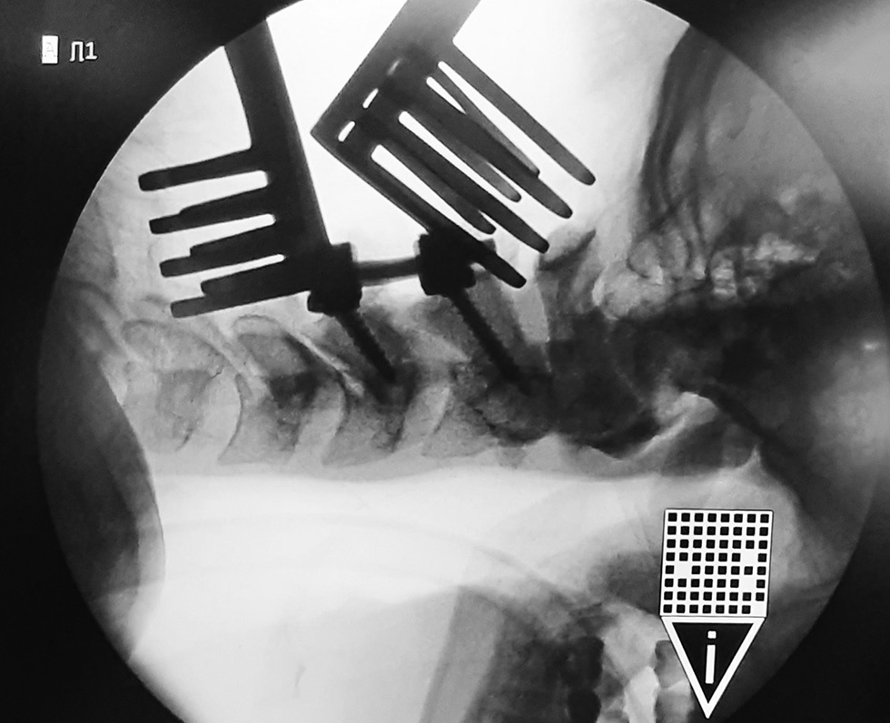
Fig. 5 Fluoroscopic image showing reduction of C2 to C3 listhesis with pedicle and pars screws in C2 and C3 vertebrae, respectively.
Fig. 5 Fluoroscopic image showing reduction of C2 to C3 listhesis with pedicle and pars screws in C2 and C3 vertebrae, respectively.
Discussion
Traumatic spondylolisthesis of the axis is caused by failure of pars interarticularis with consequent separation from the C2 vertebral body and resulting anterior slippage of the C2 over the C3 vertebra. It is typically attributed to a hyperextension or hyperflexion injury causing bending moment to the vertebra body and pedicle, causing fracture at the weakest part, that is, pars interarticularis. Burke and Harris defined atypical HF as those involving the posterior aspect of the vertebral body instead of those involving the neural arch.5 Compared with classical type, atypical fractures are also more likely to have neurological deficits. The Effendi classification, modified by Levine, is the most prevalent classification system in use for these fractures but its most important criticism is the lack of incorporation of soft tissue injury and functional component like assessment of instability.6 The longitudinal ligaments and the disk at the C2 to C3 level are the most important soft tissue elements providing stability at this level.7 However, the role of other soft tissue structure supporting the vertebrae has also been emphasized lately like posterior muscular attachments and integrity of facet joint capsules.4
The spondylolisthesis case encountered by us appears to be unusual because of the presence of C2 to C3 listhesis along with locked facet joints in the absence of any apparent bony injury to the pars, vertebral body, or pedicle. The force transferred to the cervical spine from the fall causing the instability resulting in C2 to C3 listhesis with no associated bony injury is indeed a curious case. These findings may be explained on the basis of coexisting soft tissue injuries that might not be apparent even on MRI. The operative findings reinforced and strengthened our concept of the soft tissue injury and its contribution to the instability. The muscular attachments to the posterior elements were found to be disrupted along with the interspinous and supraspinous ligaments. It has been suggested that the posterior muscular attachments would resist forward translation of vertebra.4 More importantly, integrity of the C2 to C3 facet joint capsules was also found to be compromised leading to instability. Thus, the combination of muscular injury along with loss of capsuloligamentous integrity appears to be responsible for causing the listhesis. Whether this injury represents a spectrum of atypical HF is controversial, but it shares striking similarity with the former. The component of C2 to C3 spondylolisthesis is the common factor between the two entities but with the lack of pars fracture as in our case. Instead the facet joint capsular injury provided the crucial element of instability responsible for the presentation. The importance of the facetoligamentous complex and their role in maintaining stability has been explored and established in subaxial spine; however, its role in C2 to C3 junction is still a gray area.8 9
The management algorithm for axial spondylolisthesis depends on the instability. Effendi type I and II injuries are managed conservatively, whereas operative management is favored for type IIa and III subtypes because of high pseudoarthrosis rates and poor functional outcome seen with conservative management.6 10 Although both anterior and posterior approach have been described for the treatment of this entity, the technique of posterior C2 to C3 fusion has been found to be biomechanically superior to the anterior approach.9 11 The posterior approach is further indicated in cases with locked facet joints or irreducible fractures failing closed reduction and where vertebral artery decompression is needed. This is similar to the indication in our case where we had a locked facet that could not be reduced with cervical traction. Thus, a posterior approach with open reduction of listhesis by facet joint manipulation followed by posterior fixation was the most feasible surgical approach.
Conclusion
Traumatic spondylolisthesis of the axis is a diverse condition with a wide spectrum of pathoanatomical findings in which soft tissue injury has as much a role to play as the more obvious bony injury in determining the resulting instability and subsequent therapeutic decision making. Therefore, both the factors have to be assessed and addressed to ensure successful management of this condition.
Conflict of Interest
None declared.
Funding None.
References
- On hanging, considered from a mechanical and physiological point of view. Philosophical Magazine Series 4.. 1866;32:23-34.
- [Google Scholar]
- A case of traumatic C2-3 listhesis without pars fracture: Insights from this possible variant of hangman’s fracture. Neurol India. 2017;65(1):209-211.
- [Google Scholar]
- Koller H, Kathrein A. Letter to the editor concerning: a systematic review of the management of hangman's fractures by Xin-Feng Li et al. (2006). Eur Spine J 15:257–269. Eur Spine J 2006;15(9):1415–1418., author reply 1419–1421
- A biomechanical assessment of soft-tissue damage in the cervical spine following a unilateral facet injury. J Bone Joint Surg Am. 2012;94(21):e156.
- [Google Scholar]
- In vitro genesis of subaxial cervical unilateral facet dislocations through sequential soft tissue ablation. Spine. 2001;26(12):1317-1323.
- [Google Scholar]
- A systematic review of the management of hangman’s fractures. Eur Spine J. 2006;15(3):257-269.
- [Google Scholar]
- Traumatic spondylolisthesis of the axis: a biomechanical comparison of clinically relevant anterior and posterior fusion techniques. J Neurosurg Spine. 2009;11(4):379-387.
- [Google Scholar]


