Translate this page into:
Childhood movement disorders: Clinicoetiological pattern and long-term follow-up at tertiary care center from South India
*Corresponding author: Vykuntaraju K. Gowda, Department of Pediatric Neurology, Indira Gandhi Institute of Child Health, Bengaluru, Karnataka, India. drknvraju08@gmail.com
-
Received: ,
Accepted: ,
How to cite this article: Parameshwarappa NN, Gowda VK, Shivappa SK. Childhood movement disorders: Clinicoetiological pattern and long-term follow-up at tertiary care center from South India. J Neurosci Rural Pract 2023;14:21-7.
Abstract
Objectives:
Movement disorders are common neurological problems. There is a considerable delay in the diagnosis of movement disorders which indirectly indicates their under-recognition. The studies regarding relative frequencies and their underlying etiology are limited. Describing and classifying them with a diagnosis helps in treating the condition. To study the clinical pattern of various movement disorders in children and to establish their etiology and outcome.
Materials and Methods:
This observational study was conducted in tertiary care hospital from January 2018 to June 2019. Children from 2 mo. to 18 years of age presenting with involuntary movements on the first Monday of every week were included in the study. History and clinical examination were carried out with a pre-designed proforma. A diagnostic workup was done, results were analyzed to find the common movement disorders and their etiology, and follow-up was analyzed for 3 years.
Results:
A total of 100 cases out of 158 with known etiology were included in the study of which 52% were females and 48% were males. The mean age at presentation was 3.15 years. The various movement disorders are dystonia-39(39%), choreoathetosis-29(29%), tremors-22(22%), gratification reaction-7(7%), and shuddering attacks-4(4%). Ballismus and myoclonus were found in 3(3%) children each. Tics, stereotypes, and hypokinesia were found in 2(2%) children each. A total of 113 movement disorders were found in 100 children. Etiologically, perinatal insult was the most common cause 27(27%), followed by metabolic/genetic/hereditary causes 25(25%). Infantile tremor syndrome due to Vitamin B12 deficiency-16/22(73%), was a major contributor in children with tremors. Rheumatic chorea was less in our study 5(5%). Out of the 100 study subjects, 72 cases were followed up. Out of which 26 children have completely recovered. Based on the modified Rankins score(MRS), 7 children belong to category I, 2 children belong to category II, 1 child to category III, 6 children to category IV, and 14 children to category V of MRS. A total of 16 children have died (MRS VI).
Conclusion:
Perinatal insult and Infantile tremor syndrome are more important and preventable causes. Rheumatic chorea is found to be less common. A significant number of children had more than one type of movement disorder, which warrants the need to look for varied types of movement disorders in the same child. Long-term follow-up shows complete recovery in one-fourth of children and the remaining surviving with disability.
Keywords
Pediatric movement disorder
Dystonia
Perinatal insult
Infantile tremor syndrome
INTRODUCTION
Movement disorders are characterized by impaired voluntary movement, the presence of involuntary movements, or both.[1] A movement disorder typically is defined as dysfunction in the implementation of appropriate targeting and velocity of intended movements, dysfunction of posture, the presence of abnormal involuntary movements, or the performance of normal-appearing movements at inappropriate or unintended times.[2] There is no specific diagnostic test to differentiate between movement disorders. Categorizing these movement disorders help in localizing the pathologic process, whereas the age, onset, and degree of abnormal motor activity and associated neurologic features assist in organizing the relevant investigations.[3] Movement disorders have been divided into; hyperkinetic disorders, in which there is excessive movement, and hypokinetic disorders, in which there is a paucity of movement.[1] Dyskinesia, synonymous with hyperkinetic movement disorders, includes tics, chorea, ballismus, dystonia, myoclonus, stereotypies, and tremor.[4] Hypokinetic movement disorders are sometimes referred to as akinetic/rigid disorders. The primary movement disorder in this category is Parkinsonism, primarily presenting in adulthood, and is rare in children.[2]
There are only limited studies on the frequency of movement disorders as a clinical phenomenon, their underlying etiology, and outcome.[5] There is a considerable delay in the diagnosis of movement disorders which indirectly indicate their under-recognition.[6] Laboratory investigations in this setting are directed at establishing a possible etiology underlying the child’s specific movement disorder. Determining the etiology is important for the specific treatment and prognostication. As there are limited Indian studies regarding the etiological evaluation of movement disorders, we planned this study with the objectives of studying the clinical profile of childhood movement disorders and establishing their underlying etiology and outcome.
MATERIAL AND METHODS
This prospective observational study was conducted from January 2018 to July 2022 at a tertiary-level pediatric hospital in Bengaluru, India. Children in the age group of 2 months–18 years with abnormal movements presenting at neurology, general outpatient department, and admitted patients on Monday of every week formed the study group. Children with involuntary movements suggestive of epilepsy alone were excluded from the study. Written and informed consent was taken from the parents/caregivers of children before enrollment. History and clinical examination were done with the help of a pre-designed pro forma. Any available videographic evidence of such perceived movement disorder was reviewed. The cause and type of movement disorder was evaluated based on information collected by history and clinical examination. Diagnostic workup by specific laboratory and radiological investigations such as CT and MRI of the brain, fundus examination, thyroid function tests, autoantibodies against NMDA receptor, and metabolic screen (blood gas analysis, ammonia, lactate, tandem mass spectrometry, and gas chromatography–mass spectrometry) was done as and when required. Exome sequencing by next-generation sequencing was considered when genetic etiology was suspected.
Data were entered into Microsoft Excel and analyzed using SPSS (Statistical Package for the Social Sciences, Ver.10.0.5) package. The results were averaged (mean + standard deviation) for continuous data and dichotomous data were presented in tables with numbers and percentages.
After 3 years of follow-up, study subjects were contacted through phone calls and the present disability status of the child was assessed. Modified Rankin scale was used to categorize the outcomes. Modified Rankin scale contains six categories, Category 0 is with no disability and Category 6 denotes the death of the patient. The study was approved by the Institutional Ethical Committee.
RESULTS
A total of 158 cases out of 100 cases with identified etiology were included in the study. Forty-eight (48%) were male and 52 (52%) were female. The mean age at presentation was 3.15 years. The mean duration of the disease was 12.74 months. The duration of the disease was <12 months in 74% of the children. [Table 1] shows various subtypes of movement disorder noted with etiology. [Table 2] shows the etiology of movement disorders. A total of 113 movement disorders were found in 100 children, 12 children had mixed movement disorders. Etiologically, cerebral palsy secondary to perinatal insult was the most common cause 27 (27%) followed by metabolic/genetic/hereditary causes 25 (25%). Perinatal insult was the most common cause of dystonia. Infectious/ immune-mediated cause was the most common etiology causing choreoathetosis. Approximately 72% of the tremors were caused by infantile tremor syndrome (ITS). Gratification reaction and shuddering attacks were the most common movement disorders found in the psychogenic/behavioral causes. Among the perinatal insult, hypoxic-ischemic encephalopathy (HIE) was the major category accounting for 19% of the cases followed by bilirubin-induced neurological dysfunction (BIND).
| Movement disorders | Perinatal insult | Infections/immune mediated | Metabolic/genetic/hereditary causes | Psychogenic | Miscellaneous causes |
|---|---|---|---|---|---|
| Dystonia (n=39) | HIE (16), BIND (4) | NMDAR encephalitis (2), SSPE (2), rickettsial encephalitis (1) | Leigh’s disease (6), glutaric aciduria (4), Wilson’s disease (2), organic aciduria (1), mitochondrial DNA depletion syndrome (1) | None | None |
| Choreoathetosis (n=29) | HIE (4), BIND (1) | NMDAR encephalitis (5), rheumatic chorea (5), rickettsial encephalitis (1), dengue encephalitis (1), SSPE (1) | Dihydropteridine reductase (DHPR) deficiency (1), NCL (3), Wilson’s disease (2), atypical Cockayne syndrome (1), glutaric aciduria (1), mitochondrial DNA depletion syndrome (1), neuronal migration disorder (1), organic aciduria (1) | None | None |
| Tremors (n=22) | BIND (1), stroke (1) | NMDAR encephalitis (1), post-viral cerebellitis (1), dengue cerebellitis (1) | MTHFR deficiency (1) | None | ITS (16) |
| Gratification reaction (n=7) | HIE (1), meningitis (1) | None | None | 5 | None |
| Shuddering attacks (n=4) | Meningitis (1) | None | None | 3 | None |
| Ballismus (n=3) | None | VME (1), NMDAR encephalitis (1) | Wilson’s disease (1) | None | None |
| Tics (n=2) | None | None | None | None | Tics (2) |
| Myoclonic jerks (n=3) | None | Neuroblastoma (2) | DHPR deficiency (1) | None | None |
| Stereotypies (n=2) | None | None | CDG (1) | Rett syndrome (1) | None |
| Hypokinesia (n=2) | None | None | Wilson’s disease (2) | None | None |
VME: Viral meningoencephalitis, BIND: Bilirubin-induced neurologic dysfunction, HIE: Hypoxic-ischemic encephalopathy, NHBI: Neonatal hypoglycemic brain injury, CDG: Congenital disorders of glycosylation, SSPE: Subacute sclerosing panencephalitis, ITS: Infantile tremor syndrome, MTHFR: Methylenetetrahydrofolate, NMDAR: N-methyl-D-aspartate receptor, NCL: Neuronal ceroid lipofuscinosis
| Primary category | Subcategory | Number (%) |
|---|---|---|
| Perinatal insult (n=27, 27%) | HIE | 19 (19) |
| BIND | 5 (5) | |
| Meningitis | 2 (2) | |
| Stroke | 1 (1) | |
| Infections or immune mediated (n=22, 22%) | NMDAR encephalitis | 7 (7) |
| Rheumatic chorea | 5 (5) | |
| Infections | 5 (5) | |
| SSPE | 3 (3) | |
| Neuroblastoma | 2 (2) | |
| Metabolic/genetic/hereditary causes (n=25, 25%) | Leigh’s disease | 6 (6) |
| Glutaric aciduria | 4 (4) | |
| Wilson’s disease | 4 (4) | |
| NCL | 3 (3) | |
| CDG | 1 (1) | |
| DHPR deficiency | 1 (1) | |
| Mitochondrial disorders | 1 (1) | |
| Organic aciduria | 1 (1) | |
| MTHFR deficiency | 1 (1) | |
| Neuronal migration disorder | 1 (1) | |
| Atypical Cockayne syndrome | 1 (1) | |
| Rett syndrome | 1 (1) | |
| Psychogenic or behavioral causes (n=8, 8%) | Gratification reaction | 5 (5) |
| Shuddering attacks | 3 (3) | |
| Miscellaneous causes (n=18, 18%) |
ITS | 16 (16) |
| Tic disorder | 2 (2) |
VME: Viral meningoencephalitis, BIND: Bilirubin-induced neurologic dysfunction, HIE: Hypoxic-ischemic encephalopathy,
NHBI: Neonatal hypoglycemic brain injury, CDG: Congenital disorders of glycosylation, SSPE: Subacute sclerosing panencephalitis, ITS: Infantile tremor syndrome, MTHFR: Methylenetetrahydrofolate,
NMDAR: N-methyl-D-aspartate receptor,
NCL: Neuronal ceroid lipofuscinosis
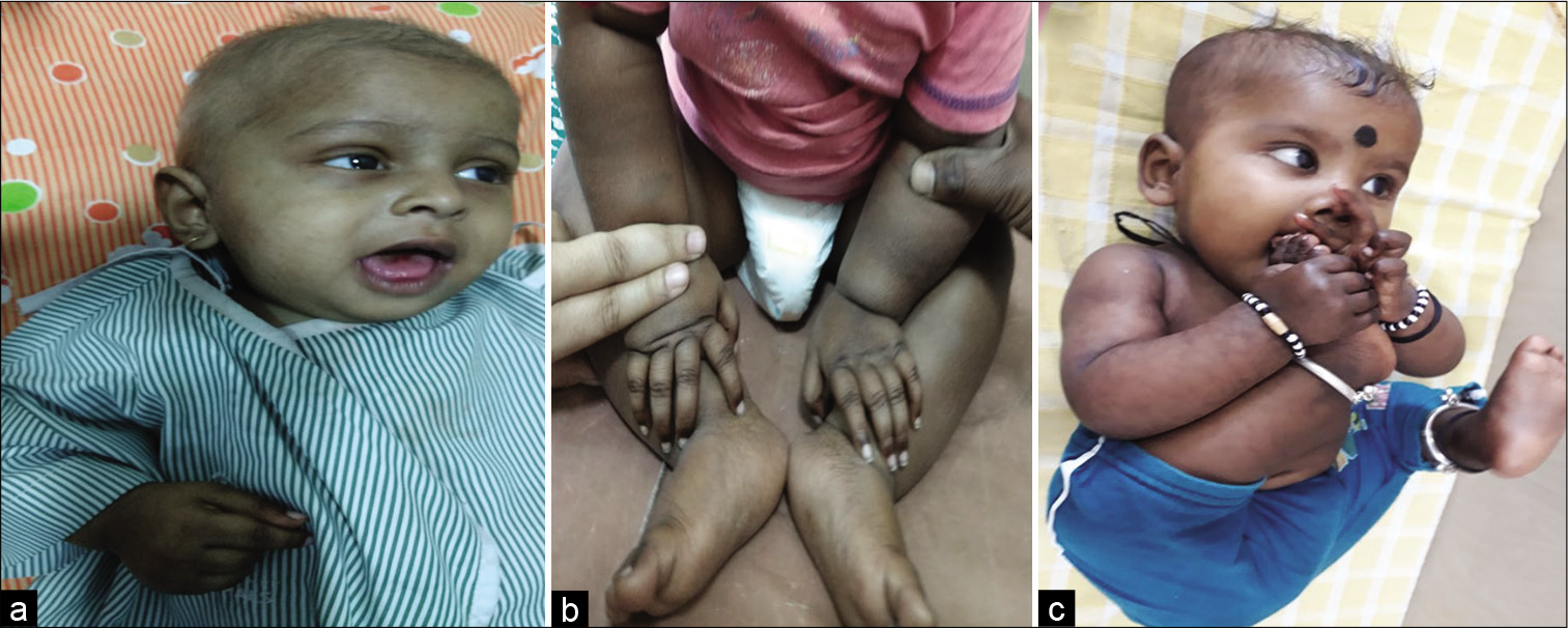
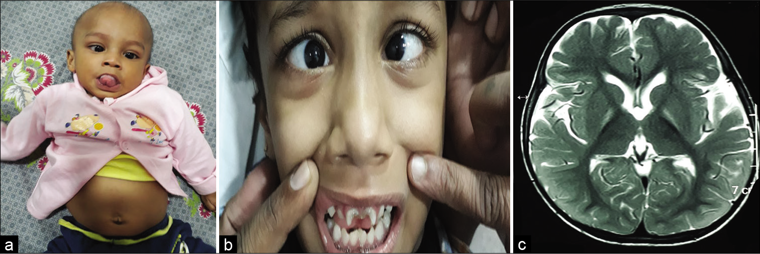
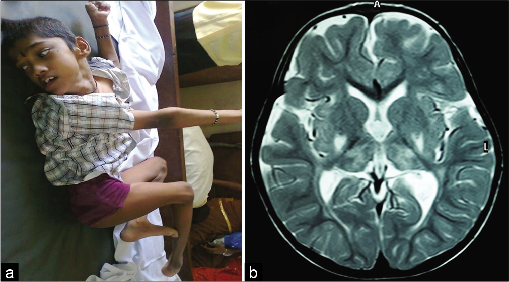
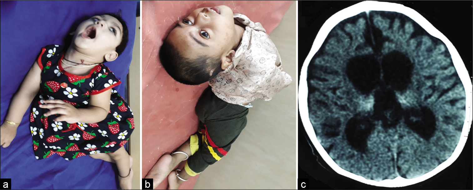
[Figure 1] shows hypopigmented (a) and sparse hair (c), hyperpigmentation of skin and knuckles (b), and apathetic child (c) in ITS. [Figure 2] shows dystonia of the tongue (a), notching in the upper incisor teeth (b), and hyperintensities in the bilateral globus pallidus in the MRI of the brain in a child with BIND. [Figure 3] shows dystonia (a) and hyperintense signal changes in the bilateral posterior putamen and thalamus in the MRI of the brain in a child with dyskinetic cerebral palsy due to birth asphyxia. [Figure 4] shows severe dystonia (a and b) and thalamic calcification (c) in the CT scan of the brain in a child with dystonic cerebral palsy due to birth asphyxia.

- Children with infantile tremor syndrome show hypopigmented (a) and sparse hair (c), hyperpigmentation of skin and knuckles (b), and apathetic child(c).

- Children with bilirubin-induced neurological dysfunction showed dystonia of the tongue (a), notching in the upper incisor teeth (b), and MRI of the brain T2W images showing hyperintensities in the bilateral globus pallidus.

- A child with dyskinetic cerebral palsy due to hypoxicischemic encephalopathy showing dystonia of limbs (a) and MRI of the brain showing hyperintense signal changes in the bilateral posterior putamen and thalamus in the T2W sequences (b).

- A child with dystonic cerebral palsy with severe arching of back and neck due to dystonia (a and b) and CT brain showing bilateral thalamic calcifications with cerebral atrophy with enlarged ventricles due to hypoxic-ischemic insult (c).
After three years of follow-up, out of the 100 cases of movement disorders, 28 cases were lost to follow-up. Other 72 cases were contacted through phone calls and the present disability status of the child was assessed. [Table 3] shows follow-up results. Out of the 72 cases, 27 children have completely recovered, seven children belong to Category I, one child each belonged to Categories II and III, six children belonged to Category IV, and 14 children belonged to Category V of modified Rankin’s score (MRS). A total of 16 children have died (MRS VI).
| Etiology | Subcategory | Modified Rankin scale (MRS) | Death (MRS-6) | Lost to follow-up | |||||
|---|---|---|---|---|---|---|---|---|---|
| 0 | 1 | 2 | 3 | 4 | 5 | ||||
| Perinatal insult (27) | HIE (19) | 2 | 6 | 3 | 8 | ||||
| BIND (5) | 1 | 2 | 2 | ||||||
| Meningitis (2) | 1 | 1 | |||||||
| Stroke (1) | 1 | ||||||||
| Infections/immune mediated (22) | Autoimmune encephalitis (anti-NMDA) (7) | 3 | 3 | 1 | |||||
| Rheumatic chorea (5) | 4 | 1 | |||||||
| Infections (5) | 1 | 2 | 2 | ||||||
| SSPE (3) | 1 | 1 | 1 | ||||||
| Neuroblastoma (2) | 1 | 1 | |||||||
| Metabolic/genetic/hereditary (25) | Leigh’s disease (6) | 1 | 2 | 3 | |||||
| Glutaric aciduria (4) | 1 | 1 | 2 | ||||||
| Neuro-Wilson’s disease (4) | 1 | 3 | |||||||
| NCL (3) | 2 | 1 | |||||||
| CDG (1) | 1 | ||||||||
| DHPR deficiency (1) | 1 | ||||||||
| MEGDEL (1) | 1 | ||||||||
| Organic aciduria (1) | 1 | ||||||||
| MTHFR deficiency (1) | 1 | ||||||||
| Neuronal migration disorder (1) | 1 | ||||||||
| Atypical Cockayne syndrome (1) | 1 | ||||||||
| Rett syndrome (1) | 1 | ||||||||
| Psychogenic/behavioral (8) | Gratification reaction (5) | 3 | 1 | 1 | |||||
| Shuddering attacks (3) | 1 | 2 | |||||||
| Miscellaneous (18) | ITS (16) | 13 | 3 | ||||||
| Tic disorder (2) | 1 | 1 | |||||||
| Total | 100 | 27 | 7 | 1 | 1 | 6 | 14 | 16 | 28 |
VME: Viral meningoencephalitis, BIND: Bilirubin-induced neurologic dysfunction, HIE: Hypoxic-ischemic encephalopathy, NHBI: Neonatal hypoglycemic brain injury, CDG: Congenital disorders of glycosylation, SSPE: Subacute sclerosing panencephalitis, ITS: Infantile tremor syndrome, MTHFR: Methylenetetrahydrofolate, NMDAR: N-methyl-D-aspartate receptor, NCL: Neuronal ceroid lipofuscinosis
Etiology-wise perinatal insult cases were 18 (out of 27), infections/immune-mediated causes included 18 (out of 22), metabolic/genetic/hereditary causes included 17 (out of 25), psychogenic/behavioral causes included five (out of eight), and the miscellaneous group included 14 (out of 18) cases.
Out of the 18 cases in the perinatal insult group, one child has recovered completely (post-meningitis), one child (post-stroke) has a persistent right-sided weakness with tremors which have reduced and are able to carry out all the daily activities, one child (kernicterus) is able to walk independently, and dystonia has reduced with medication. Two children (HIE) are requiring assistance for daily activities and for mobility. Six children (HIE) and two children (kernicterus) are bedridden requiring constant nursing care. Four children have died (three children [HIE] and one child [kernicterus]).
Out of the six cases of anti-NMDA autoimmune encephalitis, three children have completely recovered with good scholastic performance and three children have poor scholastic performance with subnormal intelligence. Out of the five cases of rheumatic chorea, four children have recovered completely; in one child, mild chorea is persistent but is able to go to school with poor scholastic performance. Out of three cases of infectious cause, one child is able to carry out all daily routine activities; other two have a moderately severe disability. Out of two cases of SSPE, one child is bedridden requiring assistance for all the activities and myoclonic jerks are persistent. One more child has died two years after the onset of the symptoms. Out of the two cases of neuroblastoma, one child had ganglioneuroma (mature type) diagnosed at the age of 18 months and underwent surgery at the same time; now, her myoclonus has reduced and she is able to sit with support, does not walk, and has good intelligence and speech. Another child was diagnosed with neuroblastoma (immature type) at the age of one year 7 months and died at 2.5 years of age even after treatment.
Out of the three cases of Leigh’s disease, one child (3.5 years old) is bedridden requiring constant nursing care, able to speak few words with good comprehension, and two children have died at age of 3 years and 3.5 years. Out of two cases of glutaric aciduria, one child diagnosed at the age of 9 months (started on treatment) is bedridden requiring constant nursing care, does not speak, and has an intellectual disability. The other child was diagnosed at the age of 10 months and started on treatment but died at the age of 1.5 years with progressive symptoms. Both the children with NCL have died (NCL-I at the age of 6.5 years and NCL-VI at the age of 3 years). One child with CDG is now 8.5 years old, able to sit with support, not able to stand, and has an intellectual disability as well. One child with 3-methylglutaconic aciduria (MEG), deafness (D), encephalopathy (E), and Leigh-like disease (L) is now 11 years old and is bedridden which requires constant nursing care and has persistent choreoathetosis. One child with MTHFR deficiency diagnosed at the age of 1 year is bedridden requiring constant nursing care and tremors are persistent even after treatment. One child with neuronal migration disorder died at the age of 15 years with progressive symptoms. One child with atypical Cockayne syndrome is now 12 years old, going to school and there are no involuntary movements at present. One child who was diagnosed with Rett syndrome at the age of 4.5 years is now 8 years old and does not speak; she can sit and walk independently but does not go to school. There were two cases of gratification reaction, which has resolved completely at 3 years of age without any medication in one child and is going to school. The other child is now 4.5 years old and still has GR which has reduced in frequency to once a day (initially 15– 20 times/day). One child with shuddering attacks has recovered completely without any medication.
Out of the 13 cases of ITS, 12 children have recovered completely. In one child who is now 5-years old, tremors have stopped but have subnormal intelligence compared to same age peers. One child was diagnosed with hot water epilepsy and tics, convulsions controlled with medication, and no tics at present.
Out of the 72 cases, 16 children have died due to various causes, the most common (15 out of 16 cases) cause of death being respiratory infections in the bedridden children except in one child where cardiorespiratory arrest after surgery for neuroblastoma was noted.
DISCUSSION
Out of 100 children, a slight female preponderance was seen with 52% being females. The mean age was 4.18 years with a range of 2 months–16 years. The mean duration of the disease was 12.74 months. [Tables 4 and 5] show a comparison of our study with the previously reported series.
| Study | Dale et al.[7] (n=52) | Goraya[5] (n=92) | Baumer et al.[6] (n=606) | Fernández-Alvarez[8] (n=964) | The present study (n=104) |
|---|---|---|---|---|---|
| Age group | 2 months–15 years | 5 days–15 years | Children and adults with childhood onset | Children and adults with childhood onset | 2 months–to 18 years |
| Mean age at presentation | 5.06 years | 4.75 years | 8 years | 5.13 years | 3.15 years |
| Males | 21 | 63 | 422 | 603 | 48 |
| Females | 31 | 29 | 184 | 361 | 52 |
| Choreoathetosis | 20 | 19 | 19 | 31 | 29 |
| Dystonia | 17 | 21 | 73 | 223 | 39 |
| Tremors | 12 | 15 | 9 | 163 | 22 |
| Myoclonus | 10 | 25 | 9 | 61 | 3 |
| Tics | Excluded | 2 | 346 | 417 | 2 |
| Ballismus | 0 | 0 | 0 | 0 | 3 |
| Hypokinesia | 10 | 3 | 13 | 13 | 2 |
| Others | 0 | 7 | 137 | 56 | 13 |
In the present study, dystonia was found to be the most common movement disorder, unlike other studies. This may be attributed to the fact that perinatal insult was included in our study, the major etiology causing dystonia; whereas other studies excluded perinatal insult. Choreoathetosis was one of the major movement disorders found in our study similar to Goraya[5] and Dale et al.[7] A study conducted by Baumer et al.,[6] including both children and adults with childhood onset of movement disorder, showed tics as the most common movement disorder followed by dystonia. This particular observation was attributed by the author as a referral bias with an over-representation of tic disorders and dystonia, given the place of study, was a special research interest in Gilles de la Tourette syndrome and dystonia, with an under-representation of other disorders, particularly tremor. The frequency of tics was relatively less in our study; this might be due to the under-recognition of mild-to-moderate cases and referral of only severe cases to our center. Fernandez et al.,[8] studied 964 cases of childhood-onset movement disorders with a mean age of 5.13years. The most common type of movement was tics, followed by dystonia, and tremors as shown in Table 4.
Out of the 29 cases of choreoathetosis, only 5 (17.24%) were attributed to rheumatic chorea. This was similar to a study conducted by Dale et al. where only 3/20 (15%) cases of choreoathetosis were caused by rheumatic chorea, whereas, in the study conducted by Goraya, 6/19 (31.58%) cases of choreoathetosis were caused by rheumatic chorea. Earlier literature showed rheumatic chorea as the most common cause of chorea in children; however, it was not the case in the present study. This may be attributed to better diagnosis as well as treatment of streptococcal infections and preventing carrier status.
Out of 22 cases of tremors, 16 (72.72%) were caused by ITS secondary to Vitamin B12 deficiency. Hence, ITS was the major etiology causing tremors in children comparable to a study conducted by Goraya where 13/15 (86%) cases were contributed by ITS. This similarity was found in our developing country, whereas there was no case of ITS in the study conducted by Dale et al. in Australia.
Hyperkinetic movement (111) disorder was far more common than hypokinetic movement disorder,[2] in keeping with all other studies compared. This might be attributed to the fact that Parkinsonism which is the major cause of bradykinesia is a rare disease in children.
In the present study, perinatal insult was the most common etiology followed by metabolic/genetic/hereditary causes. Considering single disease entity, ITS secondary to Vitamin B12 deficiency was the second most common etiology following perinatal insult, as nutritional diseases are relatively common in India. A relatively large number of cases had infectious/ immune-mediated etiology, reflecting the larger incidence of infections in tropical countries. Psychogenic/behavioral causes are relatively uncommon in our country compared to others.
Follow-up showed complete recovery in 27 out of 72 children (37.5%). The cure rate was highest in children with ITS (12 out of 13) followed by acute rheumatic chorea (four out of five) and anti-NMDA receptor autoimmune encephalitis (three out of six). In the behavioral/psychiatric disorders, four children out of five have completely recovered. Sixteen out of 72 (22.22%) children have died, the most common cause of death being lower respiratory tract infections. The limitation of the present study is that it cannot be generalized as it is done in a tertiary care referral center.
CONCLUSION
Perinatal insult was found to be the most common cause indicating the need for improved perinatal care. Infantile tremor syndrome also formed a significant part of the cohort. Rheumatic chorea is less in our study and might be attributed to the improving living status and adequate treatment of predisposing streptococcal infections. A significant number of children had more than one type of movement disorder, which warrants the need to look for varied types of movement disorders in the same child, and treatment of the same. Many movement disorders can be cured, some can be controlled to improve the quality of life of the child. And thus, early recognition and treatment are recommended.
Declaration of patient consent
Institutional Review Board (IRB) permission obtained for the study.
Conflicts of interest
There are no conflicts of interest.
Financial support and sponsorship
Nil.
References
- Movement disorders: An overview. In: Swaiman KF, Ashwal S, Ferrieri DM, Schor NF, Finkel RS, Gropman AL, eds. Swaiman's Paediatric Neurology: Principles and Practice (6th ed). Netherlands: Elsevier; 2018. p. :706-16.
- [Google Scholar]
- Movement disorders In: Kliegman RM, Stanton BF, StGeme JW, Schor NF, eds. Nelson Textbook of Pediatrics Vol 03. (1st South Asia ed.). India: Thomson Press; 2015. p. :2888-96.
- [Google Scholar]
- Common movement disorders in children: Diagnosis, pathogenesis, and management. Neurosci Med. 2012;3:90-100.
- [CrossRef] [Google Scholar]
- Acute movement disorders in children: Experience from a developing country. J Child Neurol. 2015;30:406-11.
- [CrossRef] [PubMed] [Google Scholar]
- Childhood-onset movement disorders: A clinical series of 606 cases. Mov Disord Clin Pract. 2016;4:437-40.
- [CrossRef] [PubMed] [Google Scholar]
- A prospective study of acute movement disorders in children. Dev Med Child Neurol. 2010;52:739-48.
- [CrossRef] [PubMed] [Google Scholar]
- Prevalence of paediatric movement disorders In: Fernán-dez-Alvarez E, Arzimanoglou A, Tolosa E, eds. Paediatic Movement Disorders. Paris: John Libbey Eurotext; 2005. p. :1-18.
- [Google Scholar]


