
Translate this page into:
Characteristic Vertebral Imaging in Sickle Cell Disease
Address for correspondence: Dr. Rajaraman Kartikueyan, Department of Neurosurgery, National Neurosciences Centre, Peerless Hospital Campus, 2nd Floor, 360 Panchasayar, Kolkata - 700 094, West Bengal, India. E-mail: doctorkartik2007@gmail.com
This is an open access article distributed under the terms of the Creative Commons Attribution-NonCommercial-ShareAlike 3.0 License, which allows others to remix, tweak, and build upon the work non-commercially, as long as the author is credited and the new creations are licensed under the identical terms.
This article was originally published by Medknow Publications & Media Pvt Ltd and was migrated to Scientific Scholar after the change of Publisher.
A 32-year-old woman presented with multiple episodes of self-limiting, severe back pain over several years. She was a known patient of sickle cell anemia. Magnetic resonance imaging scans of the dorsal and lumbar spines showed “H-shaped” vertebrae on coronal and sagittal imaging [Figures 1 and 2].
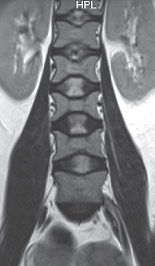
- Coronal magnetic resonance slice showing “H-shaped” lumbar vertebrae (Lincoln log vertebra) with central end plate depression consequent to infarction
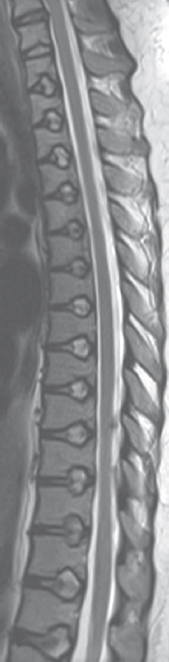
- Sagittal magnetic resonance slice (T2 sequences) showing multilevel bulging of intervertebral discs into the vertebral body
Sickle cell anemia is a condition where red blood cells (RBCs) contain abnormal hemoglobin (Hemoglobin S).[12] When deoxygenated this hemoglobin becomes insoluble and aggregates with similar molecules distorting the shape of the RBCs making them less deformable while they flow through the capillary bed.[1] The abnormally shaped RBCs also have a propensity to adhere to the endothelium.[1] All these lead to vascular occlusion and tissue infarction which manifests clinically as the painful “sickling crisis.”[12]
The microvasculature of the endplates of the vertebrae is a low flow system fed by terminal branches that arise from the arterial grid at the centrum of the vertebrae.[3] Further, the endplates themselves are usually <1 mm thick and are thinnest in the central region.[4] The combination of both these factors leads to endplate infarction with a sharply defined central depression in sickle cell disease as seen in our patient.
Financial support and sponsorship
Nil.
Conflicts of interest
There are no conflicts of interest.
REFERENCES
- The role of the vertebral end plate in low back pain. Global Spine J. 2013;3:153-64.
- [Google Scholar]
- The vertebral endplate: disc degeneration, disc regeneration. Eur Spine J. 2006;15(Suppl 3):S333-7.
- [Google Scholar]


