Translate this page into:
Airway management for occipital encephalocele in neonatal patients: A review of 17 cases
Address for correspondence: Dr. Zeynep Baysal Yιldιrιm, Department of Anesthesiology and Reanimation, Dicle University, 21280 Diyarbakir, Turkey. E-mail: zbaysal2003@yahoo.com
This is an open-access article distributed under the terms of the Creative Commons Attribution-Noncommercial-Share Alike 3.0 Unported, which permits unrestricted use, distribution, and reproduction in any medium, provided the original work is properly cited.
This article was originally published by Medknow Publications and was migrated to Scientific Scholar after the change of Publisher.
Abstract
Introduction:
Encephalocele, midline defect of cranial bone fusion, occurs most frequently in the occipital region. Airway management in pediatric patients with craniofacial disorders poses many challenges to the anesthesiologist. The purpose of this study is to describe the airway problems encountered for such cases, and describe how these problems were managed.
Materials and Methods:
We reviewed the charts of occipital encephalocele newborn that were treated by surgical correction in Harran University Hospital during 2006–2008. The collected data were categorized into preoperative, intraoperative, and postoperative data.
Results:
The mean age of the patients was 5.17 days. Of these 17 patients, eight patients (47.1%) had hydrocephaly, one patient (5.8%) with Dandy Walker syndrome. Micrognathia, macroglossia, restriction in neck movements were recorded as the reasons in six cases each. No major anesthetic complication was found.
Conclusions:
We reported perioperative management in 17 occipital encephalocele infant. Comprehensive care during peroperative period is essential for successful outcome.
Keywords
Anesthesia
difficult intubation
occipital encephalocele
Introduction
An encephalocele is cystic congenital malformation in which central nervous system structures herniated through a defect in the cranium in communication with cerebrospinal fluid pathways.[1] The incidence of encephalocele is 1 per 5000 live births.[2] The incidence of encephalocele is reported to vary in different parts of the world depending on the geographic region, gender of the affected infants, and nutritional supplementation during pregnancy, maternal age and parity. Etiology of encephalocele is still unknown.[3]
Difficult or failed tracheal intubation is feared by all anesthesiologists, and there have been many attempts to develop a means of predicting it.[4] Difficult intubation in pediatric patients is especially important because the use of predictive tests, as a part of routine clinical practice, is limited in this patient group. The occurrence of associated congenital abnormalities might be a clue for potential difficulty of intubation.
The purpose of this study is to describe the anesthetic problems encountered for these cases, and describe how these problems were managed.
Materials and Methods
After our institutional ethical committee approval, we retrospectively reviewed the records of all patients with occipital encephalocele aged between 1 and 5 days, who were treated surgically in Harran University Hospital between January 2006 and July 2008.
Hospital files of patients that had difficult intubations were evaluated for further knowledge related to difficult intubation. Demographic characteristics, associated congenital abnormalities, and perioperative airway and/or respiratory complications were also recorded.
All data were given as mean ± standard deviation (SD) with range with SPSS 11.0 (SPSS Inc, Chicago, Illinois, USA) for Windows.
Results
Data from 17 consecutive patients were analyzed. The demographic data of the patients is shown in Table 1. Of the 17 cases, 11 were female (64.7%) and 6 male (35.3 %) and ages ranged from 1 to 5 days (mean 2.1), weighted between 2700 and 3500 g (mean 3150) and head circumference is between 32 and 38 cm (mean 34). One encephalocele had two cystic sacs. Of these 17 patients, eight patients (47.1%) were hydrocephalous, one patient (5.8%) was Dandy Walker syndrome. Micrognathia, macroglossia, restriction in neck movements were recorded as the reasons of difficulty intubation in six cases each.

Anesthesia induction was done with an inhalation technique. Neuromusculer blockers were not administered. There were no ventilation problems in any of the children. In all six cases, endotracheal intubation was achieved via multiple attempts and/or maneuvers, changing blades or using stylets. Fiberoptic guidance and laryngeal mask airway could not require in any of the cases.
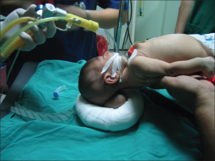
Two children were positioned in the lateral position and induced anesthesia with sevoflurane in 100% oxygen via face mask. Two attempts of direct laryngoscopy (Miller blade size 0) in the lateral position showed just the tip of the epiglottis (Cormack-Lehane Grade 3), and we could not intubate the child with this limited view. We lifted the baby off the operation table with the help of two residents [Figure 1]. One resident stabilized the child's head, and the other supported the pelvis. Laryngoscopy in this position improved visualization (Cormack-Lehane Grade 2). We intubated the trachea with a 2.5-mm uncuffed, endotracheal tube, and proceeded uneventfully.[56]

- Alternative method
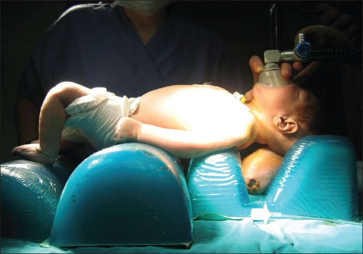
Four children were intubated with an alternative method. In this method, we used the silicone supports [Figure 2]. In our cases the silicone supports was put down on top of each other under the neonate body until the height matched that of the encephalocele sac. The neonate was put in the supine position with the body on the platform while a resident temporarily supported the head.[7]

- Mowafi method
Maintenance of anesthesia was with sevoflurane in air with O2 and opioid supplementation. Five percent glucose in 1⁄3 normal saline was administered. Blood loss was replaced by packed red cell. The duration of surgery and anesthesia for these operations was 1.35 ±0.11 h and 1.51 ± 0.19 h, respectively. No airway problems were encountered intraoperatively in our series. Hemodynamics and end-tidal carbon dioxide were stable throughout surgery in every case. We extubated all of the 17 cases (100%) in the operating room.
Discussion
This retrospective study demonstrated our 3-year experience in anesthesia in 17 patients underwent surgically repairment of occipital encephalocele.
Encephaloceles are a type of the congenital malformation seen commonly. Occipital encephaloceles represent approximately 85% of lesions seen in the western hemisphere.[1] Airway management in pediatric patients with craniofacial malformation poses many challenges to the anesthesiologist. Isada et al. reviewed the practice of anesthesia in 13,557 pediatric cases and reported that the risk of difficult intubation is higher in children with congenital malformation.[8]
Anesthetic management of these children requires carefully attention because the size of occipital encephaloceles may vary from small to large masses.[1] In this study, while one patient had a small head with a circumference of 31 cm only, another had an encephalocele with a circumference of 38 cm.
Sometimes, the occipital encephaloceles can cause to restriction of head movement. This led to difficulty in positioning for laryngoscopy and in visualizing the glottic opening. We have encountred difficult airway management in six patients having large occipital encephalocele. Hence, we tried two different methods for airway management. One of them is platform method described by Mowafi et al.[7] We applied this method to four patients. We think that this method is very useful for anesthesiologists, especially in cases of large occipital encephalocele. We used this method because this method has some advantages such as needing less resident and preventing pressure on encephalocele sac and possible rupture. Alternative approach is to lift the baby off the table,[56] which we applied to two patients. We think this method is not helpful for patients with large occipital encephalocele because it needs two residents.
Different airway management has been defined by various authors for occipitale encephalocele. Quezado et al described a simple foam-cushion devices.[9] In this approach, only one person is needed to manage the airway and pressure on the encephalocele sac is prevented.
We used sevoflurane for induction and maintenance of general anesthesia for all patients because of getting rapid induction and not being irritable.
Conclusions
We have presented here our experience in anesthesia management in patients with occipital encephalocele, who underwent surgical repair. There was no major anesthesia complication in our series. Careful perioperative management allowed us to achieve successful outcomes.
Source of Support: Nil.
Conflict of Interest: None declared.
References
- A rare case of upper airway obstruction in an infant caused by basal encephalocele complicating facial midline deformity. Paediatr Anaesth. 1999;9:73-6.
- [Google Scholar]
- Etiology, pathogenesis and prevention of neural tube defects. Congenit Anom. 2006;46:55-67.
- [Google Scholar]
- Predicting difficult intubation—Worthwhile exercise or pointless ritual? Anaesthesia. 2002;57:105-9.
- [Google Scholar]
- Neonatal airway management in occipital encephalocele. Anesth Analg. 2006;103:1632.
- [Google Scholar]
- Airway management in neonates with occipital encephalocele – adjustments and modifications. Pediatric Anesth. 2007;17:1111-21.
- [Google Scholar]
- Positioning for anesthetic induction of neonates with encephalocele? 2001. The Internet Journal of Anesthesiology. 5 Available from: http://www.ispub.com/ostia/index.php?xmlPrinter=true&xmlFilePath=journals/ija/vol5n3/enceph.xml
- [Google Scholar]
- Airway management in neonates with occipital encephalocele: Easy does it. Anesth Analg. 2008;107:1446.
- [Google Scholar]


