Translate this page into:
Endoscopic endonasal repair of spontaneous sphenoid sinus lateral wall meningocele presenting with cerebrospinal fluid leak
Address for correspondence: Dr. Ali Erdem Yildirim MD, Department of Neurosurgery, Ankara Numune Research and Education Hospital, Ankara, Turkey. E-mail: alierdemyildirim@gmail.com
This is an open-access article distributed under the terms of the Creative Commons Attribution-Noncommercial-Share Alike 3.0 Unported, which permits unrestricted use, distribution, and reproduction in any medium, provided the original work is properly cited.
This article was originally published by Medknow Publications & Media Pvt Ltd and was migrated to Scientific Scholar after the change of Publisher.
Abstract
Spontaneous sphenoid sinus lateral wall meningoceles are rare lesions with an unknown etiology. Endoscopic endonasal technique is a considerable route in the treatment of this condition. The aim of this paper is to report the etiology, surgical technique, and outcome in a patient repaired via endoscopic endonasal approach. A 51-year-old male patient applied with rhinorrhea started three months ago after an upper respiratory infection. There were no history of trauma or sinus operation. Biochemical analysis of the fluid was positive for beta-2-transferrin. This asypthomatic patient had undergone for repairment of lateral sphenoid sinus meningocele with endoscopic endonasal transsphenoidal approach. After endoscopic endonasal meningocele closure procedure no complications occured and a quick recovery was observed. Endoscopic endonasal approach is an effective and safe treatment modality of spontaneous lateral sphenoid sinus meningoceles and efficient in anterior skull base reconstruction.
Keywords
Cerebrospinal fluid leak
endoscopic endonasal surgery
meningocele
sphenoid sinus lateral wall
spontaneous
Introduction
Meningoencephaloceles or meningoceles of sphenoid sinus are infrequent lesions which occur as a result of trauma, iatrogenic injury, or skull base erosion due to inflammatory or neoplastic disorders.[1] Spontaneous meningocele of the sphenoid sinus lateral wall is extremely rare and the theory behind that is the incoplete fusion of sphenoid greater wing embriyologic precursor with presphenoid and basisiphenoid which can be concluded in a persistent channel called the lateral craniopharyngeal (Stenberg) canal.[12] Erosion of skull base and increased intracranial pressure with the background of spontaneous meningocele of lateral sphenoid sinus is another suggested theory.[3] Also in congenital skull base, defects may be diagnosed in adulthood incidentally during an investigation for headache, CSF-rinorrhea or meningitis.[1] Beta 2 transferrin, MRI and CT cisternographies should be done for diagnosis. The main target for the treatment is to prevent complications such as meningitidis, intracranial abscess and pneumocephalus.[4]
Traditional treatment for CSF-leak via craniotomy with 70-80% success.[4] Besides some known advantages of the transcranial approach, there is a 40% recurrence rate reported with the increased morbidity including anosmia, frontal lobe injury, seisures, memory deficits and intracranial hemorrages.[25] Endoscopic endonasal technique eliminates most of the limitations and became highly considerable. With the endoscopic procedure not only an excellent visualization is provided but there are also many outcome studies reporting decreased morbidity rates and improved closure rates.[6] Besides being safe and efficient, endoscopic endonasal surgery is also sufficient in protection of nasal anatomic structures and normal neural functions. In this report, we present a rarely seen case of spontaneous meningocele of lateral sphenoid sinus who received a endoscopic endonasal repair procedure.
Case Report
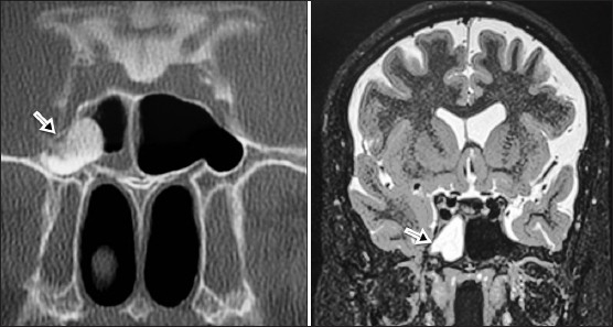
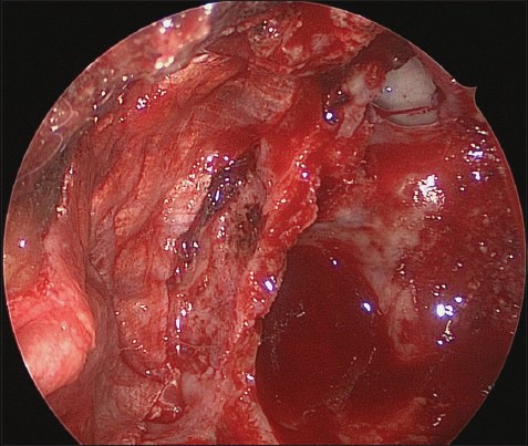
A 51-year-old male patient applied to our hospital with the complaint of rhinorrhea started three months ago after an upper respiratory infection. There were no any other symptoms present such as headache or fever and also there were no history of trauma or sinus operation. Biochemical analysis of the fluid was positive for beta-2-transferrin. Cranial MRI and CT cisternography images show a fluid collection in the right sphenoid sinus and confirm a lateral sphenoid sinus meningocele [Figure 1]. The patient was operated via endoscopic endonasal transsphenoidal approach. In the surgical procedure, a meningocele sac originating from the lateral wall of the right sphenoid sinus exposed subsequent to removal of the sphenoid sinus floor by the right mononostril approach. A CSF leakage was observed from the meningocele sac [Figure 2]. After resection of the meningocele by bipolar electrocoagulation, remnants of the sac were pushed through the bony defect in the lateral wall of the sphenoid sinus by using 30 degree optic endoscope. For a multilayer complete closure of the bony defect, we used fat and fascia lata grafts respectively, harvested from the right lateral thigh incision [Figure 3]. That followed with the application of fibrin sealent. We did not use a lumbar drainage cathether in this patient.

- Coronal cranial MRI (T2-weighted) and CT cisternography images of the patient. Arrows showing the lateral sphenoid sinus meningocele

- Endoscopic view of sphenoid sinus. Arrow showing the meningocele sac

- Endoscopic view of sphenoid sinus after meningocele repairment with fascia lata autograft
Postoperatively the patient was conscious, responsive and his general condition was good with normal vital functions. No postoperative complication occured and he was discharged at the fifth day of hospitalization. After six months of postoperative follow-up, there was no reccurrence or complication observed.
Discussion
Spontaneous meningocele of lateral sphenoid sinus is an extreme incident without a well understood etiology. Anterior skull base meningoceles could be classified based on the location as described by Van Nouhuys and Bruyn: Sphenoorbital, sphenoethmoid, transethmoid (cribiform), sphenomaxillary, and transsphenoid.[7] Medial parasellar lesions are relatively more common and may occur with the empty sella syndrome.[1] The lateral wall meningoceles of the sphenoid sinus are uncommon and generally occur in 26-40% of the patients that have extensive lateral sphenoid sinus pneumatization into the pterygoid process.[8] A defect of the middle cranial fossa may result in herniation of the temporal lobe into the sphenoid sinus and CSF leakage.[1] Congenital lateral sphenoid sinus meningoceles are exceedingly rare and theoretically arise from the developmental errors that ocur during the embryogenesis. Incomplete fusion of the greater sphenoid wings with the presphenoid and basisphenoid can result in a persistent lateral craniopharyngeal canal (Sternberg canal) which is first described by Sternberg.[2] Persistence of craniopheringeal canal may also result in a meningocele and CSF leak.[1] For the diagnosis of meningocele, various diagnostic modalities, including biochemical (beta-2 transferrin) and imaging studies (CT, MRI, and cisternography), have been previously described in the literature.[9] The treatment of patients with spontaneous meningocele of the lateral sphenoid sinus is controversial in the literature. There are many surgical repairment procedures described, but there is no common consensus in this management yet. Previously, treatment of skull base meningoceles performed by transcranial approach with a success rate of 70-80% which is not sufficient in terms of neurosurgery. Transcranial approach has some advantages but, besides its advantages, it causes various important morbidities like anosmia, frontal lobe retraction, seizure induction, memory loss and intracranial bleeding.[5] Today, with advancement of endoscopic surgery, it is possible to treat these diseases with endoscopic endonasal transsphenoidal approach which is a relatively noninvasive technique. There are many reports supported endoscopic approaches to this area.[610] The use of both angled scopes and excellent visualization without any brain retraction is the main advantages of this technique. We obtained a favorable result in our spontaneous lateral sphenoid sinüs meningocele case by conducting endoscopic endonasal repair procedure.
Spontaneous sphenoid sinus lateral wall meningoceles may occur because of increased ICP or persistant lateral craniopharyngeal canal. The clinical manifestations of these lesions are used to be CSF rhinorrhea, headache, and meningitis. By the transsphenoidal approach it is easier to access these lesions located in the sphenoid sinus lateral recess then the transcranial way and the usage of endoscope in endonasal transsphenoidal approach provides excellent visualization and high successful closure rates.
Source of Support: Nil.
Conflict of Interest: None declared.
References
- Endoscopic management of spontaneous meningoencephalocele of the lateral sphenoid sinus. J Neurosurg. 2010;112:1070-7.
- [Google Scholar]
- Sternberg's canal cause of congenital sphenoidal meningocele. Eur Arch Otorhinolaryngol. 2000;257:430-2.
- [Google Scholar]
- Intrasphenoidal transsellar encephalocele repaired by endoscopic approach. Ann Otol Rhinol Laryngol. 2003;112:890-3.
- [Google Scholar]
- Endoscopic endonasal repair of anterior skull base non-traumatic cerebrospinal fluid leaks, meningoceles, and encephaloceles. J Neurosurg. 2010;113:961-6.
- [Google Scholar]
- Cerebrospinal fluid fistula: Clinical aspects, techniques of localization, and methods of closure. J Neurosurg. 1969;30:399-405.
- [Google Scholar]
- Transnasal endoscopic repair of cerebrospinal fluid rhinorrhea: A meta-analysis. Laryngoscope. 2000;110:1166-72.
- [Google Scholar]
- Nasopharyngeal transsphenoidal encephalocele, craterlike hole in the optic disc and agenesis of the corpus callosum. Pneumoencephalographic visualization in a case. Psychiatr Neurol Neurochir. 1964;67:243-58.
- [Google Scholar]
- The importance of the lateral extensions of the sphenoid sinus in post-traumatic cerebrospinal rhinorrhea and meningitis. J Neurosurg. 1965;22:326-32.
- [Google Scholar]
- Cerebrospinal fluid rhinorrhea: Diagnosis and management. Otolaryngol Clin North Am. 2005;38:597-611.
- [Google Scholar]
- Endonasal endoscopic repair of Sternberg's canal cerebrospinal fluid leaks. Laryngoscope. 2007;117:345-9.
- [Google Scholar]


