Translate this page into:
Orbital extension of trigeminal schwannoma
Address for correspondence: Dr. Shantanu Ghosh, C/4/49 Kendriya Vihar, Kolkata - 700 052, West Bengal, India. E-mail: shashwata_shantanu@yahoo.co.in
This is an open-access article distributed under the terms of the Creative Commons Attribution-Noncommercial-Share Alike 3.0 Unported, which permits unrestricted use, distribution, and reproduction in any medium, provided the original work is properly cited.
This article was originally published by Medknow Publications & Media Pvt Ltd and was migrated to Scientific Scholar after the change of Publisher.
Abstract
Schwannomas, also known as neurilemmomas, are benign peripheral nerve sheath tumors. Trigeminal schwannomas are rare intracranial tumors. Here, we report a 35-year-old female presenting with an axial proptosis of right eyeball with right-sided III, IV and VI cranial nerve palsy. Her best corrected visual acuity in the right eye was perception of light absent and in the left eye was 20/20. MRI scan revealed a large right-sided heterogeneous, extra-axial middle cranial fossa mass that extended to the intraconal space of right orbit. A diagnosis of intracranial trigeminal nerve schwannoma with right orbital extension was made. Successful surgical excision of the mass with preservation of the surrounding tissues and orbital exenteration was done. Post-operative period was uneventful.
Keywords
Extracranial extension
orbit
trigeminal schwannoma
Introduction
Schwannomas are usually benign tumors arising from the schwann cells in the axon myelin sheaths.[1] Trigeminal schwannomas are uncommon intracranial tumors, accounting for 0.8% to 8% of intracranial schwannomas. Trigeminal schwannomas are much less common than acoustic neuroma.[2] This benign tumor grow larger and spread into extracranial compartments through the foramina in the skull.[3] The variable location and presentation of this tumor makes the diagnosis difficult. The diagnosis is usually confirmed by histopathological examination. Here we present a rare case of benign intracranial trigeminal schwannoma arising from right parasellar region extending to intraconal space of the right orbit which was managed successfully by surgery.
Case Report
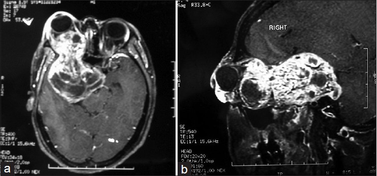
A 35-year-old female presented with a painless, progressive diminution of vision in right eye since 3 years. She also complained of forward protrusion of right eyeball for past 2 years. General physical examination was unremarkable. On ocular examination best corrected visual acuity in the right eye was perception of light absent and in the left eye was 20/20. Exophthalmometry showed 3 mm proptosis in the right eye. The size of proptosis did not vary with posture or with valsalva maneuver. The restricted ocular movements in all gazes in the right eye were suggestive of right-sided III, IV and VI cranial nerve palsy. The ocular movements in the left eye were normal. There was diminished corneal sensation in the right eye. The rest of the cranial nerve examinations were normal. Slit lamp examinations of the eyes showed dilated pupil in the right side which was not reacting to light. The fundus examination revealed secondary optic atrophy in the right eye and normal left eye. Hematological tests were within normal limits. MRI images of brain and orbit [Figure 1a and b] demonstrated a large well-marginated extra-axial middle cranial fossa mass with myxomatous regions at the right anterior temporal region extending into right intraconal space of orbit through optic foramen and superior orbital fissure to produce displacement of optic nerve and eyeball to cause proptosis. The mass was hypointense on T1, hyperintense with hypointense septae on T2 and marked enhancement of the mass with diffuse irregular hypointensity on hyperintense background on gadolinium-enhanced sequences were suggestive of trigeminal schwannoma. There was encasement of the right internal carotid artery. The mass extended into right suprasellar area, interpeduncular fossa, perimesencephalic and cerebropontine angle cistern to cause contralateral shifting of the brain stem. CT of the brain revealed destruction of lesser wing and body of sphenoid bone on the right side with enlargement and erosion of ipsilateral superior orbital fissure, foramen ovale and foramen rotundum by the large hypodense well-marginated densely enhancing mass. The tumor entered the orbit through the superior orbital fissure which was extraconal. To become intraconal in the orbit, the tumor had eroded the lesser wing of sphenoid between the superior orbital fissure and the optic foramen thereby entering the annulus of Zinn. On the basis of the MRI and CT features and the location of the mass, a suggestive diagnosis of schwannoma of ophthalmic division of right trigeminal nerve was made. The patient was taken up for an excision biopsy under general anesthesia. A right-sided pterional craniotomy with orbitozygomatic extension was done. The mass was found to be extradural in position covered on the surface by the outer layer of the dura. This was dissected off and piece meal excision of the tumor mass was done including the intraorbital portion due to attachments to adjacent structures. The distal portions of right-sided III, IV and VI cranial nerves from the tumor could hardly be recognized intraoperatively. The right eyeball was enucleated by the ophthalmologist and the tumor masses were sent for histopathological examination.
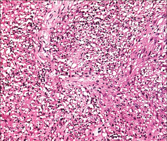
The postoperative period was uneventful except mild numbness in the right half of the forehead. On histopathological examination, a biphasic pattern of tumor cells were seen with areas of closely packed spindle cells having fusiform nuclei and eosinophilic cytoplasm (Antoni A). In some places cells were arranged in palisades referred to as Verocay bodies. There was no evidence of malignant transformation. All these features confirmed the diagnosis of benign schwannoma [Figure 2]. After about 6 month follow up there were no signs of recurrence of the tumor.

- (a and b) Axial and sagittal MRI scan of brain and orbit showing a large well-marginated extra-axial middle cranial fossa mass with areas of cystic degeneration extending into the orbit

- Histopathology showing neoplastic spindle cells arranged in fascicles and whorls (H and E stain, ×100)
Discussion
Trigeminal schwannomas are slow-growing encapsulated tumors composed of differentiated schwann cells of sensory root. It can originate in any section of the trigeminal nerve. Trigeminal schwannomas are classified in four types according to the locations: Middle fossa type (type A), posterior fossa root type (type B) where the tumor is in front of brainstem, dumbbell-shaped type with both middle and posterior fossa components (type C) and extracranial tumor with intracranial extension (type D).[45] Yoshida and Kawase classified extracranial schwannomas into infratemporal, orbital and pterygopalatine fossa components.[6] The schwannoma of orbit accounts for 1-4% of all orbital neoplasm.[7] The tumor has no significant sex predilection and age of presentation varies from 6 months to 72 years. Because of the subtle nature of symptoms and neglect of early symptoms, majority of the tumors achieve a large size before their diagnosis is made. The variable origin and location of this tumor causes combination of signs and symptoms in the patients.[8] Paresthesia or numbness in more than one division of the nerve are common complaints of patients rather than severe neuralgic pain. Wasting of the temporalis and pterygoid muscles are usually diagnostic for the disorder. The corneal reflex is often diminished or absent. Larger size of the tumor can involve the adjoining cranial nerves in the cavernous sinus. The larger tumor is responsible for relatively uncommon symptoms of increased intracranial pressure and ophthalmoscopically demonstrated papilledema. Radiologically the erosion of the petrous apex and erosion of foramen ovale, rotendum, superior orbital fissure and displacement of the internal carotid artery are characteristic and have diagnostic value. About 24% of orbital schwannomas have been reported to originate from the first division of the trigeminal nerve. The displacement of the eyeball is related to the site and extent of the tumor mass. The orbital tumor can present with proptosis, diplopia, limitation of eye ball movement and neuropathy. In our case most of those features were present. Most schwannomas require early treatment as they grow progressively. The growth of the tumor may compress the optic nerve to produce optic disc edema or optic atrophy. The tumor should be removed earliest to prevent optic nerve compression.[9] This benign tumor may undergo malignant change. Trigeminal schwannomas are removed by relatively small and straightforward exposures with minimum brain handling. Surgical excision of the tumor is the treatment of choice and radiation therapy is an alternative treatment in some cases. Incomplete excisions may lead to recurrences.
In our case, the tumor was a trigeminal schwannoma affecting the first division of the trigeminal nerve (Ophthalmic division). There were also III, IV and VI cranial nerve palsy of right side causing restricted extraocular movements in the right eye. She had optic atrophy due to compression of the optic nerve by the tumor.
A schwannoma though rare should be considered as a differential diagnosis of a unilateral slow growing orbital mass particularly in an adult and early intervention is needed to prevent development of vision-threatening complications.
Source of Support: Nil.
Conflict of Interest: None declared.
References
- Trigeminal neuroma: Analysis of surgical experience with 73 cases. Neurosurgery. 2003;52:783-90.
- [Google Scholar]
- Magnetic Resonance Imaging of the Brain and Spine (2nd ed). Philadelphia: Lippincott Raven; 1996. p. :781-6.
- The trigeminal schwannomas with some remarks on malignant invasion of the gasserian ganglion. Clin Neurosurg. 1955;1:11-54.
- [Google Scholar]
- Trigeminal neurinomas extending into multiple fossae: Surgical methods and review of the literature. J Neurosurg. 1999;91:202-11.
- [Google Scholar]


