Translate this page into:
Congenital spinal intramedullary epidermoid cyst
*Corresponding author: Dr. Abhinav Chander Bhagat, Department of Radiodiagnosis, All India Institute of Medical Sciences, Bhopal, Madhya Pradesh, India. abhinav.bhagat811@gmail.com
-
Received: ,
Accepted: ,
How to cite this article: Sreekanth PV, Neeli H, Bhagat AC, Malik R, Meena M. Congenital spinal intramedullary epidermoid cyst. J Neurosci Rural Pract. 2025;16:128-30. doi: 10.25259/JNRP_330_2024
Abstract
Spinal epidermoid cysts are rare benign tumors, accounting for <1% of all spinal tumors, with intramedullary cases being particularly uncommon. We present a rare case of a congenital intramedullary epidermoid cyst in a 12-year-old male, who presented with progressive bilateral lower limb weakness and gait disturbance over five months. Magnetic resonance imaging revealed a well-defined intramedullary lesion at the D6-D8 level, exhibiting hypointensity on T1-weighted images, hyperintensity on T2-weighted images, diffusion restriction on diffusion-weighted images, and peripheral rim-like enhancement on post-contrast imaging, suggestive of an epidermoid cyst. Surgical resection was performed, and histopathological analysis confirmed the presence of lamellated keratinous material, consistent with an epidermoid cyst. This case is notable for the absence of associated spinal dysraphism, making it a rare presentation. Imaging features, including characteristic signal patterns and diffusion restriction, aided in the diagnosis, distinguishing it from other intramedullary lesions such as arachnoid cysts, dermoid tumors, and neoplasms.
Keywords
Calcium
Child
Febrile
Magnesium
Seizure
INTRODUCTION
Spinal epidermoid cysts are benign rare tumors comprising <1% of all spinal tumors. Most of them are intradural extramedullary in location, with intramedullary location being extremely rare. True intramedullary epidermoid cysts occurring without spinal dysraphism or prior surgery are even more infrequent, comprising 0.8% of all spinal epidermoid tumors.[1] Spinal epidermoid cyst can be congenital, frequently associated with spinal dysraphism or segmentation anomalies; or acquired in patients who have had a prior history of trauma, surgery, or lumbar puncture. These are mainly seen in children. We describe a case of a 12-year-old male with intraspinal epidermoid cyst who had no history of prior surgery or lumbar puncture and no associated spinal malformation which makes it an uncommon presentation.
CASE REPORT
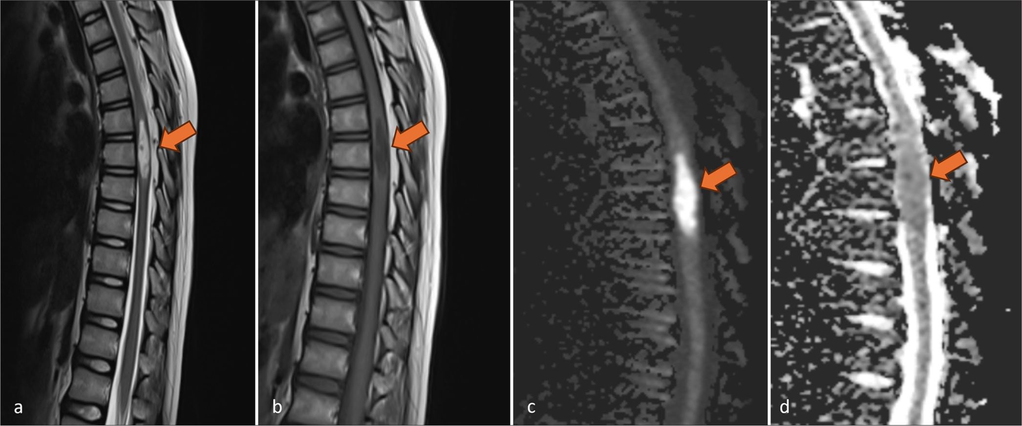
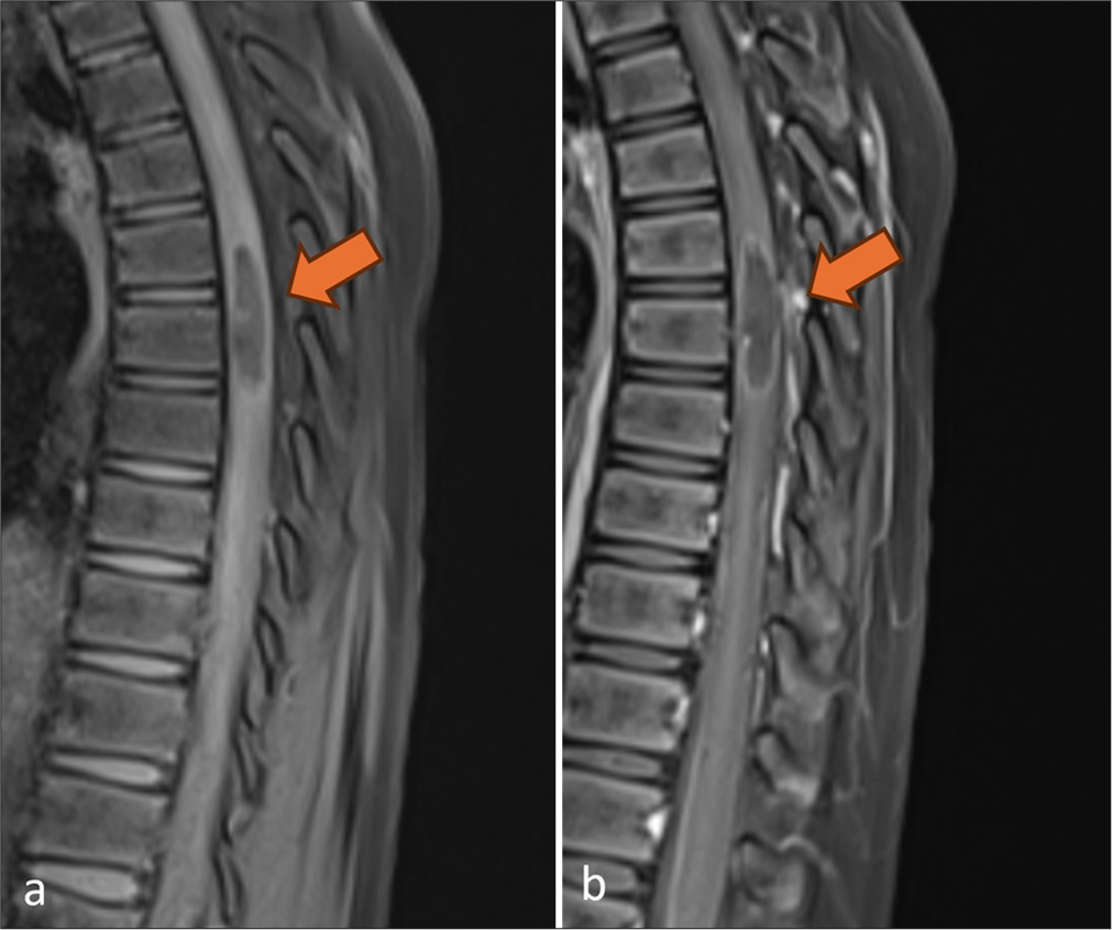
A 12-year-old male child presented with a history of insidious onset, gradually progressive bilateral lower-limb weakness for five months with a change in gait, difficulty in walking, and inability to stand up from a sitting position. No history of fever and seizures was present. There was no significant birth history or past intensive care unit admission. On physical examination, decreased power (4/5) was noted in bilateral lower limbs with intact sensory system. Magnetic resonance imaging (MRI) of the whole spine was performed which revealed a central well-circumscribed elliptical T1-hypointense and T2-hyperintense intramedullary lesion measuring approximately 0.7 × 1 × 2.9 cm (anteroposterior × transverse × craniocaudal) in maximum orthogonal dimensions at D6-D8 vertebral level causing focal symmetrical cord expansion [Figure 1]. It showed diffusion restriction with no evidence of blooming foci on the gradient-recalled echo sequence. On post-contrast study, showed mild peripheral rim-like enhancement with a central non-enhancing component [Figure 2]. No evidence of cord edema or meningeal enhancement was noted. Imaging findings were suggestive of a benign intramedullary epidermoid cyst.

- Sagittal non-enhanced magnetic resonance images of dorsal spine reveal a well-defined elliptical lesion (arrows) in dorsal cord, focally expanding the cord. It shows (a) hyperintense signal on T2-weighted images and (b) hypointense signal on T1-weighted images. (c) On diffusion-weighted imaging and (d) corresponding apparent diffusion coefficient map, the lesion shows diffusion restriction.

- Sagittal (a) pre-contrast and (b) post-contrast T1 fat-suppressed magnetic resonance images show mild peripheral rim enhancement (arrows) of the lesion.
Therapeutic intervention
The patient underwent surgical resection of the lesion which upon histopathological evaluation revealed lamellated keratinous material, consistent with an epidermoid cyst. No evidence of atypia or malignancy was noted.
Follow-up and outcome
The patient was recalled after one week, one month, and three months post-surgery for follow-up and reported improved gait and neurological status after surgery.
DISCUSSION
Spinal epidermoid cysts are rare lesions that are usually seen in children, accounting for <1% of all spinal tumors.[2] Fewer than 100 cases of intramedullary spinal epidermoid cysts have been reported. Sporadic cases of intramedullary spinal epidermoid cysts have been reported in the literature, with one case report describing cervical intramedullary location with liquid contents.[3-7] Pathogenesis of spinal epidermoid cysts can be congenital or acquired. Since both neural tube and skin develop from ectoderm during embryogenesis, epidermal cells may grow within the thecal sac. Congenital epidermoid cysts are usually associated with spinal dysraphism or segmentation anomalies such as spina bifida, myelomeningocele, diastematomyelia, syringomyelia, and hemivertebra. Acquired causes include lumbar puncture, trauma, and prior surgery. In a case series done by Morita et al., they found that out of four patients diagnosed with epidermoid cysts, three patients had a history of prior lumbar puncture.[8]
Most of the cases are asymptomatic and are incidentally detected. However, if symptomatic, clinical presentation usually depends on the location of the tumor. Congenital intramedullary epidermoid cysts are usually located in the thoracic cord and present with motor or sensory complaints in the lower limbs.[9] Acquired epidermoid cysts are usually located in the lumbar spine, likely secondary to faulty lumbar puncture, and the patients present with complaints of lower back pain. Therefore, the presentation of these tumors would be dependent on their location.
Contrast-enhanced MRI is the imaging modality of choice, where intramedullary epidermoid cysts usually appear as well-defined lesions expanding the cord, showing a hypointense signal on T1-weighted images and hyperintense signal on T2-weighted images with restricted diffusion on diffusion-weighted imaging/apparent diffusion coefficient. Depending on the internal contents, the signal characteristics may vary from iso-hyperintense on T1- and T2-weighted images. In post-contrast studies, they usually show no enhancement or may show peripheral rim-like enhancement.[10] Calcifications are rare. These lesions may also show contiguous extramedullary extension into the dural sac. Intraoperatively, intramedullary epidermoid cysts appear as encapsulated pearly white lesions.[1] Intradural extramedullary epidermoid cysts may have adhesions with arachnoid membrane making it difficult to excise completely, thereby increasing the recurrence rate. Histologically, they consist of stratified squamous epithelium with internal laminated keratin material.[11]
In this case, a 12-year-old male presented with insidious onset, gradually progressive bilateral lower limb weakness for five months with no prior history of lumbar puncture or trauma. Our case is rather rare owing to the presence of a congenital intramedullary epidermoid cyst without any associated spinal dysraphism, segmentation anomalies, or dermal sinus tract. Another close differential diagnosis considered in our case was an arachnoid cyst; however, in view of diffusion restriction and mild peripheral enhancement, it was unlikely and ruled out. Other intramedullary lesions such as dermoid and intramedullary neoplasms (astrocytoma and ependymoma) can be differentiated by the presence of fat in the former and heterogeneous enhancement of the lesion in the latter.
CONCLUSION
Spinal intramedullary epidermoid cysts are very rare spinal tumors that should be considered in children and young adolescents with motor weakness or in adults with a past history of lumbar puncture or prior trauma. An MRI plays an important role in the diagnosis of these lesions, displaying typical imaging characteristics. Imaging wise they have few differential diagnoses, with characteristic imaging and histological appearances making their diagnosis possible.
Ethical approval
Institutional Review Board approval is not required.
Declaration of patient consent
The authors certify that they have obtained all appropriate patient consent.
Conflicts of interest
There are no conflicts of interest.
Use of artificial intelligence (AI)-assisted technology for manuscript preparation
The authors confirm that there was no use of artificial intelligence (AI)-assisted technology for assisting in the writing or editing of the manuscript and no images were manipulated using AI.
Financial support and sponsorship: Nil.
References
- Spinal intramedullary epidermoid cysts: Three case presentations and literature review. Surg Neurol Int. 2020;11:17.
- [CrossRef] [PubMed] [Google Scholar]
- Cervical intramedullary epidermoid cyst with liquid contents. Asian Spine J. 2011;5:59.
- [CrossRef] [PubMed] [Google Scholar]
- Intramedullary epidermoid cyst. A case report. Neurosurg Rev. 1994;17:89-93.
- [CrossRef] [PubMed] [Google Scholar]
- Intramedullary spinal epidermoid cyst-a rare cause of spastic paraparesis. Asian J Neurosurg. 2024;19:309-11.
- [CrossRef] [PubMed] [Google Scholar]
- Epidermoid intramedullary cyst: A rare case report. J Med Cases. 2019;10:67-71.
- [CrossRef] [Google Scholar]
- Intramedullary spinal epidermoid cyst of the cervicodorsal region: A rare entity. J Pediatr Neurosci. 2010;5:49-51.
- [CrossRef] [PubMed] [Google Scholar]
- Intraspinal epidermoid tumor of the cauda equina region: Seven cases and a review of the literature. J Spinal Disord Tech. 2012;25:292-8.
- [CrossRef] [PubMed] [Google Scholar]
- A congenital spinal epidermoid cyst. J Belg Soc Radiol. 2015;99:127-8.
- [CrossRef] [PubMed] [Google Scholar]
- Spinal epidermoid tumors: Case report and review of the literature. Neurospine. 2018;15:117-22.
- [CrossRef] [PubMed] [Google Scholar]
- Evolution of epidermoid cyst into dermoid cyst: Embryological explanation and radiological-pathological correlation. Neuroradiol J. 2019;32:92-7.
- [CrossRef] [PubMed] [Google Scholar]


