
Translate this page into:
Vaginal extrusion of a ventriculo-peritoneal shunt catheter in an adult
Address for correspondence: Dr. Johnathan A. Engh, Department of Neurological Surgery, University of Pittsburgh Medical Center, 200 Lothrop Street, Suite B 400, Pittsburgh, Pennsylvania - 15213, USA. E-mail: enghja@upmc.edu
This is an open-access article distributed under the terms of the Creative Commons Attribution-Noncommercial-Share Alike 3.0 Unported, which permits unrestricted use, distribution, and reproduction in any medium, provided the original work is properly cited.
This article was originally published by Medknow Publications & Media Pvt Ltd and was migrated to Scientific Scholar after the change of Publisher.
Abstract
Ventriculo-peritoneal shunts (VPS) are commonly used in the treatment of various neurosurgical conditions, including hydrocephalus and pseudotumor cerebri. We report only the second case of vaginal extrusion of a VPS catheter in an adult, and the first case with a modern VPS silastic peritoneal catheter. A 45-year-old female with a history of VPS for pseudotumor cerebri, Behcet's syndrome, and hysterectomy presented to our institution with the chief complaint of tubing protruding from her vagina after urination. On gynecologic examination, the patient was found to have approximately 15 cm of VPS catheter protruding from her vaginal apex. A computed tomography scan of the abdomen and shunt X-ray series demonstrated no breaks in the tubing, but also confirmed the finding of the VPS catheter extruding through the vaginal cuff into the vagina. The patient had the VPS removed and an external ventricular drain was placed for temporary cerebrospinal fluid diversion. Ventricular catheter cultures were positive for diphtheroids. After an appropriate course of antibiotics, a contralateral ventriculo-pleural shunt was placed one week later. Although vary rare, vaginal extrusion can occur in adults, even with modern VPS catheters.
Keywords
Adult female
hydrocephalus
vaginal extrusion
ventriculo-peritoneal shunt

Introduction
Ventriculo-peritoneal shunts (VPS) are commonly used in the treatment of various neurosurgical conditions, including hydrocephalus and pseudotumor cerebri. Intraabdominal complications, including organ perforation, are not uncommon, and have been reported.[1] Transvaginal protrusion of a VPS catheter is exceedingly unusual, and has been reported just once in the literature in an adult with a Raimondi catheter (PubMed search indexed using key words “ventriculo-peritoneal shunt, adult, protrusion, vagina” with four other reports in children aged under one year).[2] About 40 years later, we report only the second case of vaginal extrusion of a VPS catheter in an adult, and the first case with a modern VPS silastic peritoneal catheter.
Case Report
A 45-year-old female with Behcet's syndrome presented with the chief complaint of shunt tubing protruding from her vagina after urination. The patient stated that approximately 1 month earlier, she had an episode of urinary incontinence and was treated for a urinary tract infection. Over the past 2 weeks, she has also complained of scant urinary leakage. The patient denied other symptoms, such as headache, neurologic changes, abdominal pain, or fever. Her past medical history was significant for a VPS insertion in 2001 for pseudotumor cerebri and a complete revision 13 months before presentation due to a Proprionibacterium acnes infection of the VPS. At that time, a general surgeon placed the distal shunt catheter into the abdomen using laparoscopic visualization, and a postoperative abdominal X-ray [Figure 1] was obtained for confirmation. Further history included a total abdominal hysterectomy in 2003 for endometriosis and a gastric bypass in 2004. Prior to the hysterectomy, the patient had three full-term spontaneous vaginal deliveries.
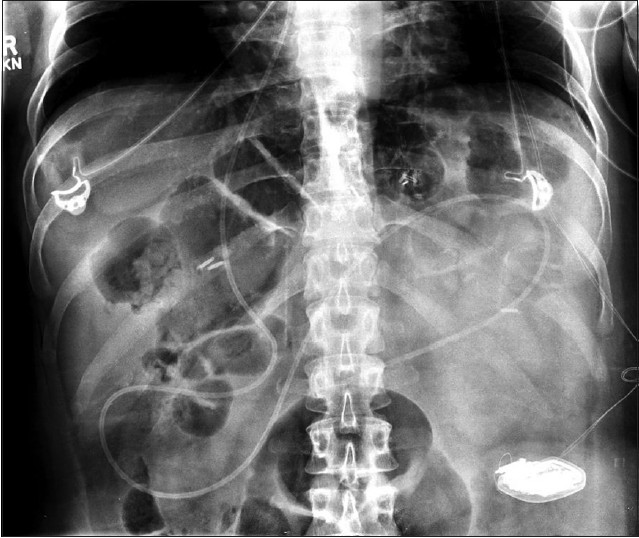
- AP X-ray illustrating the distal VPS catheter in the peritoneum
On gynecologic examination, the patient was found to have approximately 15 cm of VPS catheter protruding from her vaginal apex. The patient had no other significant physical or neurological examination findings. An abdominal X-ray [Figure 2] found no discontinuity in the tubing, but also confirmed the finding of the VPS catheter coursing through the abdomen and out of the vagina. A computed tomography scan of the head revealed no change in ventricular size or catheter location.
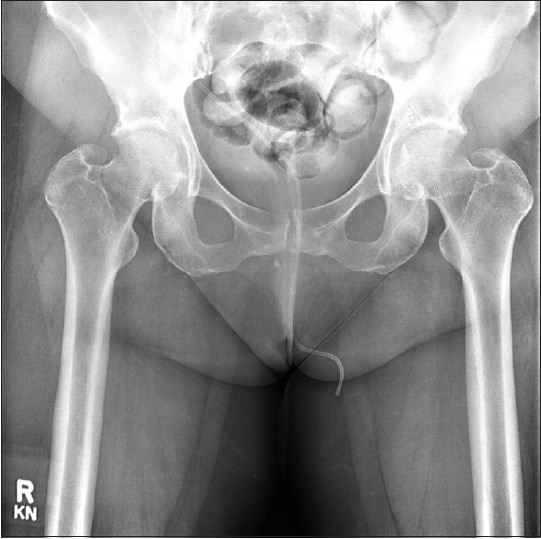
- AP X-ray illustrating the VPS catheter coursing through the pelvis and out the vagina
The patient was taken to the operating room where the cranial incisions were opened. A cystoscopy was performed that revealed an intact bladder with no evidence of any erosion of the shunt into the bladder, nor concern for any fistula formation. The vaginal cuff was grossly intact with the VP shunt coming directly through the central portion of the cuff. The VPS was sectioned distal to the valve and the peritoneal catheter was pulled out through the vagina without difficulty. The small opening in the vaginal apex was closed primarily using a 3.0 polysorb suture. The ventricular catheter was removed from the head, and an external ventricular drain was inserted. Cultures from the operating room revealed no growth in the cerebrospinal fluid (CSF) and diphtheroids from the catheter tip (RBC-1264, WBC-0, Glucose-54, Protein-15). A definitive shunt was not placed during this operation because of the patient's history of shunt infections and we wanted to monitor serial cultures to rule out a P. acnes infection. One week later, a new shunt was inserted, with the distal end placed into the pleural cavity. The patient was discharged from the hospital on 2 weeks of empiric antibiotic and antifungal coverage.
At 1 month follow-up, patient was doing well from both a gynecological and neurosurgical standpoint; however, her cranial incision did show signs of superficial breakdown. At 3 months, aside from two pinpoint scabs, her wounds had healed completely and she had no gynecological complications. Given her history of Behcet's syndrome and chronic wound healing problems she will be monitored closely.
Discussion
VPS are very common procedures used in conditions such as hydrocephalus and pseudotumor cerebri, and in cases that necessitate CSF diversion. Numerous complications involving abdominal organ perforations have been previously reported in the literature. Peritoneal catheters have protruded though the anus (after perforating the bowel), the urethra (after perforating the bladder), the mouth (after perforating the stomach), and less commonly the heart, scrotum, and lung.[345678] The majority of these cases were in infants, who presumably have weaker tissue and may be more susceptible to erosion of a foreign body.
Mozingo and Cauthen reported the first case of a VPS catheter protruding from a vagina in an adult in 1974.[2] In that case, the woman was 50 years old, and had also undergone a hysterectomy years prior. The VPS was only in place approximately 3½ months before the catheter was noticed in her vagina. The extrusion site was in the vaginal apex and the VPS used a Raimondi catheter. Our case shares characteristics with the prior report. First, both patients had previous hysterectomies and the site of extrusion was through the vaginal cuff. Despite the hysterectomies being remote and well-healed, it is likely that this area was unable to withstand chronic compression from a foreign body. Also, both extrusions happened in a relatively brief time after the offending VPS insertion (13 months in our patient, 3 months in the previous report). If prior surgery or anastomosis sites are inherently less resistant to foreign body pressure, one must wonder if patients with other intraabdominal surgeries (bowel resection, gastric bypass, etc.) are at increased risk for organ perforation as well. Other mechanisms of protrusion discussed in the literature initially include catheter fixation and anchoring to the organ serosa from a local inflammatory reaction. Subsequently, bowel peristaltic movements, increased intraabdominal pressure, and CSF pulsations are believed to provide a continuous source of mechanical pressure that ultimately lead to necrosis at the anchoring site, perforation, and finally extrusion.[9]
The presence of diphtheroids in the culture specimen from the ventricular catheter tip, which was kept separate from the abdominal-pelvic portion of the shunt tubing, may have contributed to the vaginal extrusion as well. Infections and hardware contamination are documented to contribute to wound erosion in multiple compartments of the body. However, the CSF analysis showed no evidence of inflammation, so it is unclear whether this germ was contributing to the vaginal erosion, or if it was perhaps a contaminant in this case.
The Raimondi catheters used in past VPS had a firm tip to avoid kinking. There was concern that this characteristic could contribute to organ perforation. However, in our case, the distal end of the Strata catheter (Medtronic, Inc. Minneapolis, MN) was soft and not reinforced. Thus, it appears that even with the modern silastic VPS catheters, vaginal extrusion can occur in an adult.
Managing such a complication requires a multi-disciplinary team, including neurosurgeons and gynecologists. Most importantly for repair of the protrusion, a cystoscopy must be performed to prove the bladder is not damaged. Finally, before pulling the catheter out, a stitch must be placed to keep the defect in site and then tied down once the catheter has been retrieved.
Behcet's syndrome is an immune-mediated small-vessel vasculitis that disrupts mucosal membranes as well as ocular, gastrointestinal, pulmonary, and genitourinary dysfunction.[10] Wound healing is known to be generally impaired in this patient population. While it is unknown what caused the vaginal perforation in this particular case, a history of prior hysterectomy and chronic mucosal dysfunction may have put this patient at higher risk.
Although vary rare, vaginal extrusion can occur in patients other than children. To this end, consideration of alternative placement of the distal shunt catheter outside of the abdomen may be given in patients with a history of prior hysterectomy, gynecologic surgery, or chronic mucosal immunosuppression. Finally, if the peritoneum is ultimately used as the location for placement of the distal catheter, utilizing a shorter catheter length may be beneficial.
Source of Support: Nil.
Conflict of Interest: None declared.
References
- Abdominal complications of ventriculoperitoneal shunts with emphasis on the role of imaging methods. Surg Gynecol Obstet. 1983;156:473-8.
- [Google Scholar]
- Vaginal perforation by a raimondi peritoneal catheter in an adult. Surg Neurol. 1974;2:195-6.
- [Google Scholar]
- Bowel perforation and transanal protrusion of a ventriculoperitoneal shunt catheter. Pediatr Neurosurg. 2006;42:129-31.
- [Google Scholar]
- Bladder calculus formation and urinary retention secondary to perforation of a normal bladder by a ventriculoperitoneal shunt. Urology. 2002;60:344.
- [Google Scholar]
- Migration of distal ventriculoperitoneal shunt catheter into the heart. Case report and review of the literature. J Neurosurg. 2004;100(Suppl Pediatrics 2):206-11.
- [Google Scholar]
- Perforation of the bowel complicating peritoneal shunt for hydrocephalus. Report of two cases. Am Surg. 1966;32:601-3.
- [Google Scholar]
- The incidence of inguinal complications after ventriculoperitoneal shunt for hydrocephalus. Childs Nerv Syst. 2005;21:44-7.
- [Google Scholar]
- Spontaneous extrusion of the abdominal tube through the umbilicus complicating peritoneal shunt for hydrocephalus. Case report. J Neurosurg. 1973;38:758-60.
- [Google Scholar]
- Urethral extrusion of a peritoneal catheter in a patient with a neobladder: A rare complication of shunt insertion. Neuropediatrics. 2011;42:124-7.
- [Google Scholar]
- Autoinflammatory diseases as cause of wound healing defects. Hautarzt. 2011;62:524-33.
- [Google Scholar]


