Generic selectors
Exact matches only
Search in title
Search in content
Post Type Selectors
Search in posts
Search in pages
Filter by Categories
Book Review
Brief Report
Case Letter
Case Report
Case Series
Commentary
Current Issue
Editorial
Erratum
Guest Editorial
Images
Images in Neurology
Images in Neuroscience
Images in Neurosciences
Letter to Editor
Letter to the Editor
Letters to Editor
Letters to the Editor
Media and News
None
Notice of Retraction
Obituary
Original Article
Point of View
Position Paper
Review Article
Short Communication
Systematic Review
Systematic Review Article
Technical Note
Techniques in Neurosurgery
View/Download PDF

Translate this page into:

Images in Neurosciences
11 (
1
); 211-211
doi:
10.1055/s-0039-3401965

Tuberous Sclerosis Complex with Lung Involvement

Department of Service de Neurochirurgie, Hôpital du Valais, Centre Hospitalier du Valais Romand (CHVR), Hôpital de Sion, Sion, Switzerland
Address for correspondence Arthur Robert Kurzbuch, MD Department of Service de Neurochirurgie Hôpital du Valais, Centre Hospitalier du Valais Romand (CHVR), Hôpital de Sion, Avenue du Grand-Champsec 80, CH-1951 Sion Switzerland kurzbuch@web.de
Licence
This is an open access article published by Thieme under the terms of the Creative Commons Attribution-NonDerivative-NonCommercial License, permitting copying and reproduction so long as the original work is given appropriate credit. Contents may not be used for commercial purposes, or adapted, remixed, transformed or built upon. (https://creativecommons.org/licenses/by-nc-nd/4.0/)
Disclaimer:
This article was originally published by Thieme Medical and Scientific Publishers Private Ltd. and was migrated to Scientific Scholar after the change of Publisher.

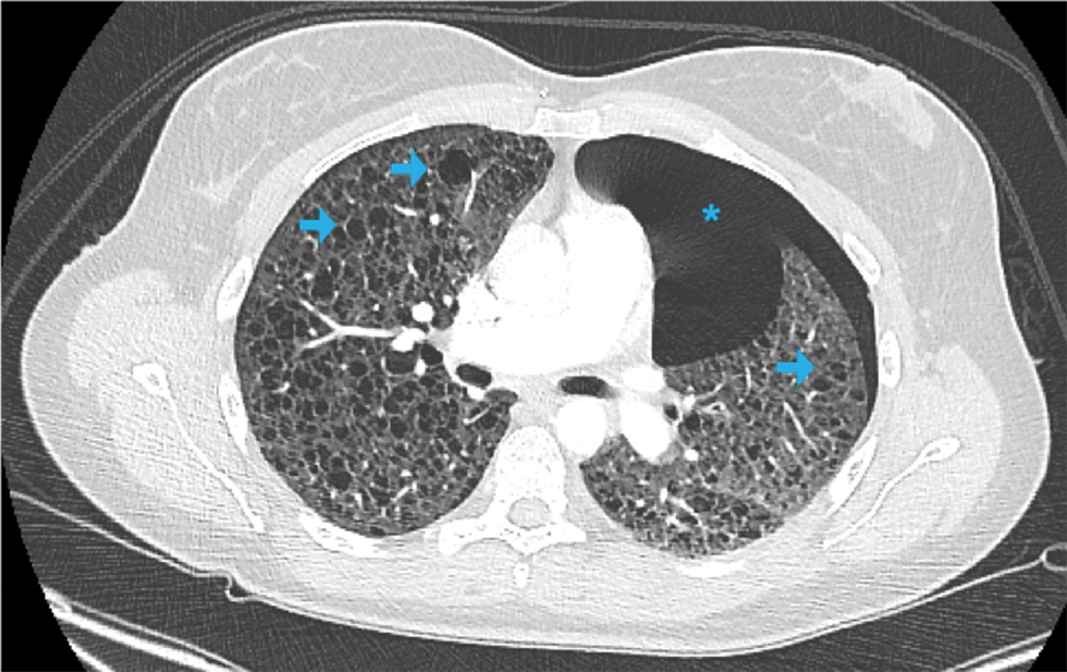
A 31-year-old woman, nonsmoker, known for tuberous sclerosis complex (TSC) underwent uneventful endoscopic resection of a growing subependymal giant cell astrocytoma localized in the left lateral ventricle. Two days after the operation the patient developed dyspnea. Computed tomography of the chest revealed a left-sided pneumothorax (blue asterix) and diffuse cystic changes throughout the lung parenchyma (blue arrows) consistent with pulmonary lymphangioleiomyomatosis (LAM; Fig. 1). TSC is an autosomal dominant disorder, resulting from mutations in chromosome 9 (TSC1 encoding hamartin) or chromosome 16 (TSC2 encoding tuberin), that manifests with multisystem involvement: skin, brain, heart, kidneys, and lungs. LAM affects predominantly women and is characterized by proliferation of abnormal smooth-muscle cells in the lung parenchyma that are responsible for cystic changes. Clinically, patients present with dyspnea and pneumothorax. The pneumothorax required draining for 4 days and the patient was discharged home 2 days later without further complications.

Fig. 1 Axial computed tomography of the chest shows diffuse cystic changes throughout the lung parenchyma consistent with pulmonary lymphangioleiomyomatosis (blue arrows) and a left-sided pneumothorax (blue asterisks).

Fig. 1 Axial computed tomography of the chest shows diffuse cystic changes throughout the lung parenchyma consistent with pulmonary lymphangioleiomyomatosis (blue arrows) and a left-sided pneumothorax (blue asterisks).

Conflict of Interest

None declared.

References

  1. , , , , , . The genetics of pneumothorax. Am J Respir Crit Care Med. 2019;199(11):1344-1357.
    [Google Scholar]
  2. , , . Pulmonary manifestations in tuberous sclerosis complex. Am J Med Genet C Semin Med Genet. 2018;178(3):326-337.
    [Google Scholar]
  3. , , , , , . Tuberous sclerosis complex. Nat Rev Dis Primers. 2016;2:16035.
    [Google Scholar]

Fulltext Views
403

PDF downloads
115
View/Download PDF
Download Citations
BibTeX
RIS
Show Sections