Translate this page into:
Transient global amnesia
Address for correspondence: Dr. Venkatraman Indiran, Department of Radiodiagnosis, Sree Balaji Medical College and Hospital, 7 Works Road, Chromepet, Chennai, Tamil Nadu, India. E-mail: ivraman31@gmail.com
This is an open access article distributed under the terms of the Creative Commons Attribution-NonCommercial-ShareAlike 3.0 License, which allows others to remix, tweak, and build upon the work non-commercially, as long as the author is credited and the new creations are licensed under the identical terms.
This article was originally published by Medknow Publications & Media Pvt Ltd and was migrated to Scientific Scholar after the change of Publisher.
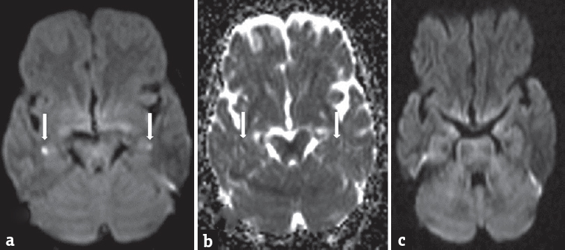
A 65-year-old afebrile female patient was brought to magnetic resonance imaging (MRI) suite, with a history of memory loss for the past 8 h. She spoke fluently but could not recognize her family members and kept asking about her whereabouts. Her neurological examination was otherwise unremarkable. She did not have a history of epilepsy, stroke, similar past episodes, hypertension, dyslipidemia, diabetes, or smoking. Laboratory testing (blood counts, biochemistry, and electrolytes) and electrocardiogram were normal. MRI showed punctuate nearly symmetrical hyperintense foci showing restricted diffusion in the bilateral hippocampal region on diffusion-weighted imaging (DWI) [Figure 1a and b]. Rest of the sequences showed no demonstrable findings. Carotid Doppler and echocardiogram were unremarkable. She recovered spontaneously 24 h after onset of clinical symptoms but was not able to remember events during the period. Diagnosis of transient global amnesia (TGA) was considered. MRI with DWI repeated after 10 days showed no abnormality [Figure 1c].

- Axial diffusion-weighted magnetic resonance imaging image (diffusion-weighted imaging) showed punctuate nearly symmetrical hyperintense foci showing restricted diffusion in the bilateral hippocampal region (a and b). Diffusion-weighted imaging repeated after 10 days showed no abnormality (c)
TGA is characterized by acute onset reversible memory disturbances (usually within 24 h) without alteration of consciousness or personal identity. Possible hypothesis includes ischemia, epilepsy, migraine, and emotional stress.[1] DWI usually shows restricted punctate foci in CA1 region in the hippocampus.[12] Abnormalities in DWI are highly variable in TGA cases (0%–84%), but it has a benign outcome and does not require any treatment.[1] However, restricted punctate foci in CA1 region in the hippocampus in appropriate clinical setting of anterograde amnesia are a very good pointer toward TGA.
Financial support and sponsorship
Nil.
Conflicts of interest
There are no conflicts of interest.
REFERENCES
- Diffusion-weighted imaging for the differential diagnosis of disorders affecting the hippocampus. Cerebrovasc Dis. 2012;33:104-15.
- [Google Scholar]


