
Translate this page into:
Third ventricular cavernoma: Report of two cases
This is an open-access article distributed under the terms of the Creative Commons Attribution-Noncommercial-Share Alike 3.0 Unported, which permits unrestricted use, distribution, and reproduction in any medium, provided the original work is properly cited.
This article was originally published by Medknow Publications & Media Pvt Ltd and was migrated to Scientific Scholar after the change of Publisher.
Sir/Madam,
Cerebral parenchyma is the most common site for a cavernoma in the nervous system. Intraventricular cavernomas are rare and constitute 2.5-10.8% of cerebral cavernomas.[1] Third ventricular cavernomas are even rare, only 25 cases have been reported in the literature.[2] We report two more cases of third ventricular cavernomas.
The first patient was a 54-year-old man, who presented with acute headache and vomiting. He was unconscious on arrival in casualty. CT scan showed third ventricular hemorrhage [Figure 1a]. He underwent external ventricular drainage, followed by ventriculo-peritoneal (VP) shunt later. An MRI done at 10 days after ictus showed a mixed density lesion with hemorrhagic component occupying the third ventricle with broad base on the floor [Figure 1b]. He underwent tranacallosal-interhemispheric approach and total excision of the lesion. The tumor was arising from the floor of the third ventricle. Histopathology was cavernoma. After surgery the patient developed diabetes insipidus, which was managed with desmopressin.
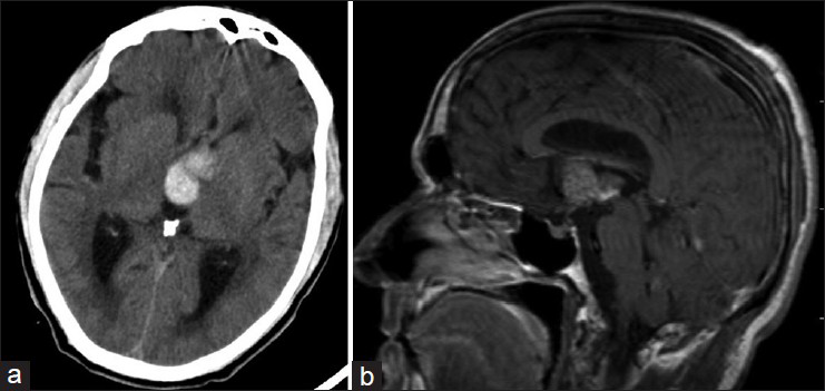
- (a) CT scan showing hemorrhage in third ventricle. (b) MRI showing a heterogeneous lesion in the third ventricle with bleed
The second patient was a 6-year-old girl who presented with headache and vomiting for 2 months. CT scan showed a calcified in third ventricle with hydrocephalus [Figure 2a]. MRI showed a heterogeneous lesion occupying the entire third ventricle [Figure 2b]. There was no evidence of recent hemorrhage. She underwent VP shunt followed by subfrontal approach, and only partial excision of the lesion as the lesion was densely adherent to the floor of the third ventricle. Histopathology was cavernoma. After surgery she developed transient hyponatremia, which was managed medically.
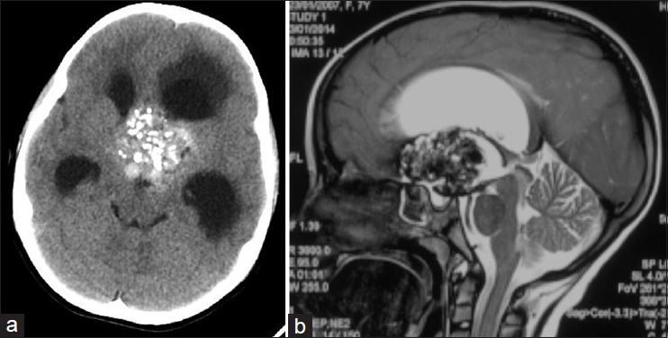
- (a) CT scan showing calcified tumor in third ventricle. (b) MRI showing a heterogeneous lesion in the third ventricle
Third ventricular cavernomas can present with signs and symptoms of any third ventricular tumors, however they usually present with symptoms of hydrocephalus.[2] Presentation as intraventriuclar hemorrhage is uncommon, and only three cases have been reported.[2] Our first patient presented with hemorrhage, and second with hydrocephalus. They can arise from the suprachiasmatic region, foramen of Monroe, lateral wall or floor of third ventricle.[3] Both of our patients had cavernomas arising from floor of third ventricle. The imaging appearance is that of typical cavernomas. The close differential diagnosis in the absence of hemorrhage is third ventricular craniopharyngioma.
The natural history of intraventricular cavernoma is not known. There is a risk of hemorrhage and acute hydrocephalus if the lesion is not excised completely. However, there is risk of hypothalamic damage in case of complete resection of lesion arising from floor of third ventricle, as it happened in our first case. It may be worthwhile doing only a VP shunt for hydrocephalus if the lesion is arising from the floor of the third ventricle.[4]
References
- Intraventricular cerebral cavernomas: A series of 12 patients and review of the literature. J Neurosurg. 2010;112:140-9.
- [Google Scholar]
- Third ventricular cavernous malformation: An unusual lesion. Br J Neurosurg. 2014;28:110-2.
- [Google Scholar]
- Intraventricular cavernomas: Three cases and review of the literature. Neurosurgery. 1999;44:648-55.
- [Google Scholar]
- Intraventricular cavernous hemangiomas located at the foramen of monro. J Korean Neurosurg Soc. 2012;52:144-7.
- [Google Scholar]


