
Translate this page into:
Serpentine calcification: A radiological stigma
This is an open-access article distributed under the terms of the Creative Commons Attribution-Noncommercial-Share Alike 3.0 Unported, which permits unrestricted use, distribution, and reproduction in any medium, provided the original work is properly cited.
This article was originally published by Medknow Publications and was migrated to Scientific Scholar after the change of Publisher.
Sir,
A 50-year-old female laborer from a low socioeconomic status came to the neurology outpatient with complaints of back pain. Her past medical history was unremarkable. There was no history of trauma and drug intake. She has been having pain in low back since last year. Pain was diffuse, mild with no radiation and parasthesia. On examination, there was no tenderness; straight leg raising test was 75° in both lower limbs. Power was grade 5/5, reflexes 2+ and plantar flexor. Sensory system examination was normal. Routine biochemistry was not contributory.
Plain radiograph of the lumbar spine view showed two soft tissue calcification in the paraspinal muscles extending from L2-5 vertebra [Figure 1]. The calcification was long, linear-beaded and fragmented in appearance. A diagnosis of guinea worm calcification was made. Patient was reassured and maintained on symptomatic treatment. She is asymptomatic on 1 year follow-up.
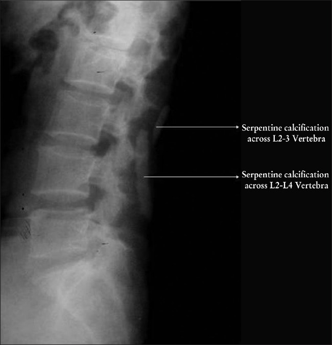
- Plain radiograph of the lumbar spine view showed two soft tissue calcification in the paraspinal muscles extending from L2-5 vertebra.
Dracunculosis or guinea worm disease, a preventable infection caused by the parasite Dracunculosis Medinensis (DM) is transmitted by drinking water containing copepods (water fleas) that are infected with DM larvae. The worm survives the digestive enzyme in the stomach and after 1 year the female migrates to the surface of the body producing a blister on the skin. If the worm does not reach the skin at all, it disintegrates or becomes calcified which can be seen on the X-ray. The muscle movement causes breaking up of the calcified worm rendering a beaded appearance.
The localization and the characteristic appearance of the calcifications in the patient were diagnostic of guinea worm calcification Guinea worm calcification may take several forms. Typically it calcifies in the lower extremities, where it assumes a long string like serpenginous or curvilinear calcification which may be very long extending to even meters. These worms are elongated, nodular, beaded and fragmented due to muscular action breaking up the worm in several areas, particularly in the calf. However, the calcification can also be oval as a result of dystrophic calcification. The causes of soft tissue calcification are 1. dystrophic calcification, 2. metastatic calcification, 3. calcinosis, 4. chondrocalcinosis, 5. synovial chondromatosis.
Dystrophic calcification could be due to venous, infection, neoplasm, drugs; vitamin D overdose, autoimmune, trauma. Venous calcification occurs within the valves, phleboliths and has a characteristic central lucency. Subcutaneous sheet like calcification are seen in postmenopausal females in venous calcification. In cysticercosis, patients have multiple “rice grain” calcifications which are oriented along the direction of the muscle fibers. Metastatic osteosarcoma and primary soft tissue osteosarcoma can be amorphous, fluffy around the bone.
All calcification have a characteristic appearance which helps us to narrow down the differential. Guinea worm calcification can attain various forms which may range from linear elongated to curvilinear to oval forms. Our patient had a linear calcification and as her calcification was in the lumbar area the size was smaller than the usual forms. Also most have a beaded appearance as was present in our case. Linear and beaded appearance and the length of the calcification present in our case have a differential of guinea worm calcification.
In the recent years DM has been eradicated from several countries.[1] But the stigma of the disease can still be seen in some patients. But this calcification was not responsible for her pain and was a coincidental finding. A high index of suspicion in endemic areas will help us in avoiding expensive investigations.


