
Translate this page into:
Segmental Neurofibromatosis
Prasad Krishnan, MS, MCh Department of Neurosurgery, National Neurosciences Centre Peerless Hospital Campus, 2nd Floor, 360, Panchasayar, Garia 700094, Kolkata, West Bengal India prasad.krishnan@rediffmail.com
This article was originally published by Thieme Medical and Scientific Publishers Pvt. Ltd. and was migrated to Scientific Scholar after the change of Publisher.
Abstract
Abstract
Segmental neurofibromas are the rarest variant of neurofibromatosis. We describe one such case in a 34-year-old man with multiple subcutaneous swellings in the posterior aspect of the left lower limb.
Keywords
mosaicism
neurofibromatosis
sectorial neurofibromatosis
segmental neurofibromatosis

Introduction
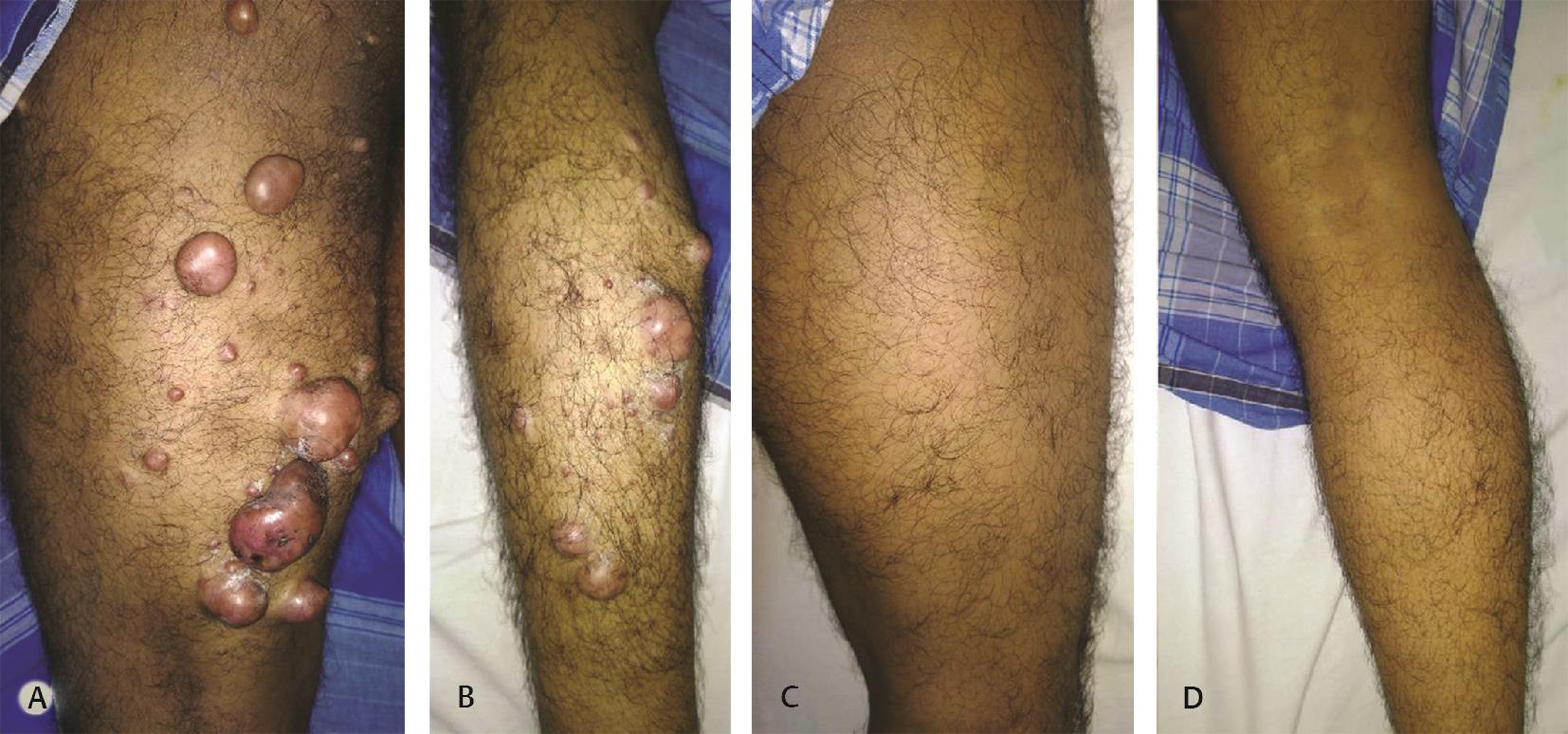
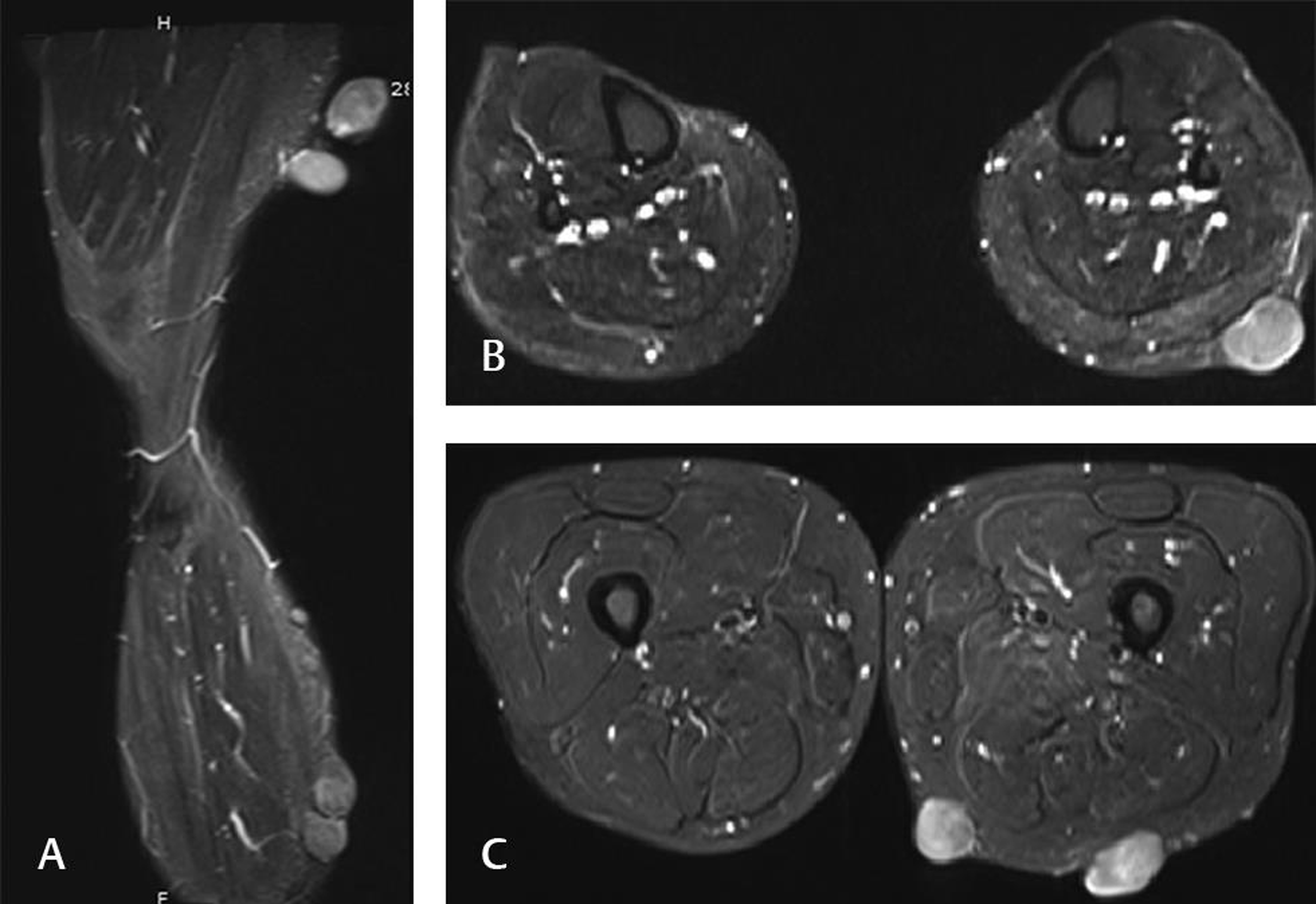
A 34-year-old male presented with multiple swellings in his left lower limb of several years duration some of which were progressively increasing in size. The swellings were only on the posterior aspect of his thigh and calf (Fig. 1). Some were tender and he had difficulty in sitting on hard surfaces. Magnetic resonance imaging (MRI) of the lower limbs revealed all of these to be subcutaneous with no deeper spread (Fig. 2). MRI of his spine and lumbar and sacral plexuses were normal. Excision biopsies of the largest ones were performed and reported as neurofibromas. Subsequently, investigations did not reveal any ocular or skeletal abnormalities or skin discolorations anywhere in the body, and further no family history for a similar disease was elicitable.
-
Fig. 1 Clinical photograph of (A) back of left thigh and (B) back of left leg of the patient showing multiple subcutaneous neurofibromas in contrast to (C) and (D) clinical photograph of back of right thigh and right lower leg which appear normal.
Fig. 1 Clinical photograph of (A) back of left thigh and (B) back of left leg of the patient showing multiple subcutaneous neurofibromas in contrast to (C) and (D) clinical photograph of back of right thigh and right lower leg which appear normal.
-
Fig. 2 Proton density fat saturated magnetic resonance imaging sequences (A) parasagittal left leg, (B) axial at level of both legs, and (C) axial at level of both thighs showing subcutaneous neurofibromas on the left side with no deeper involvement.
Fig. 2 Proton density fat saturated magnetic resonance imaging sequences (A) parasagittal left leg, (B) axial at level of both legs, and (C) axial at level of both thighs showing subcutaneous neurofibromas on the left side with no deeper involvement.
Discussion
Segmental neurofibromatosis (SNF) was first described by Crowe in 1956 and termed as sectorial neurofibromatosis.1 It was later renamed as SNF by Miller and Sparkes in 1977.1 While neurofibromatosis (NF) is among the commonest congenital diseases (1 in 3,000 births),2 3 SNF is extremely rare and its estimated prevalence is between 0.0014 and 0.002%.2 3 It is hypothesized to occur due to a postzygotic mutation of the NF1 gene late in the course of embryonic development affecting a subset of cells (localized neural crest lines) in the fetus rather than a germline NF1 gene mutation that would affect all cells in the body.4 As a consequence, the disease is localized (i.e., segmental) and further these mutations are detected only in cells in the affected region and not elsewhere in the body or in leucocytes in circulating blood as in most cases of NF.4 5
SNF was initially classified as NF type V and was considered to have the following characteristics, skin changes or neurofibromas in a single, unilateral body part with no crossing of the midline and absence of family history and systemic involvement.3 6 However, rarely bilateral SNF has been noted (6% cases)2 3 and patients with SNF have had children with systemic NF as well.3 This is explained by gonadal mosaicism.3 6 To resolve these discrepancies, Roth et al7 further classified SNF into four subtypes: true segmental, localized with deep involvement, hereditary, and bilateral. Clinically, SNFs may be divided into those with cutaneous markers alone, those with neurofibromas alone (most common) and those with cutaneous markers and neurofibromas, and those with plexiform neurofibromas.1 2 3 6
Differential diagnosis like nevus lipomatosus cutaneous superficialis, agminated lentiginosis (when only pigmentary skin changes are present), granuloma annulare, multiple fibromas, and eruptive xanthomas have been mentioned in literature6 8 but can easily be ruled out by histopathological diagnosis of an excised mass.
While there is no definitive treatment for SNF, large disfiguring and painful lesions can be treated by surgical excision.
Conflict of Interest
None declared.
Funding None.
References
- Segmental neurofibromatosis: a report of 3 cases. Indian J Dermatol. 2010;55(1):105-108.
- [Google Scholar]
- Independent NF1 mutations underlie café-au-lait macule development in a woman with segmental NF1. Neurol Genet. 2018;4(4):e261.
- [Google Scholar]
- Segmental neurofibromatosis is caused by somatic mutation of the neurofibromatosis type 1 (NF1) gene. Eur J Hum Genet. 2000;8(6):455-459.
- [Google Scholar]
- Multi-segmental neurofibromatosis. Indian J Dermatol Venereol Leprol. 2004;70(6):361-363.
- [Google Scholar]


