Translate this page into:
Giant plexiform neurofibroma presenting with craniovertebral junction instability
Address for correspondence: Dr. Bindu Menon, Professor, Department of Neurology, Narayana Medical College and Superspeciality Hospital, Chintareddypalem,Nellore-2, Andhra Pradesh, India. E-mail: bneuro_5@rediffmail.com
This is an open-access article distributed under the terms of the Creative Commons Attribution-Noncommercial-Share Alike 3.0 Unported, which permits unrestricted use, distribution, and reproduction in any medium, provided the original work is properly cited.
This article was originally published by Medknow Publications and was migrated to Scientific Scholar after the change of Publisher.
Sir,
We report a case of giant plexiform neurofibroma (PNF), presenting with progressive quadriparesis with neurofibromatosis 1 (NF1).
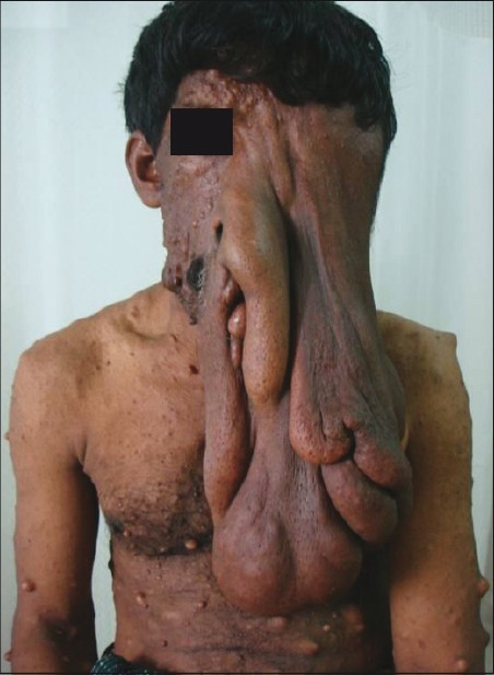
A 37-year-old male presented with gradually progressive spastic quadriparesis with bladder involvement since the last 1 year. The patient had a massive irregular peduntulous mass extending from the left forehead to the mid abdomen [Figure 1] with multiple neurofibromas all over the body. The mass was lobulated, firm, nontender, and it entirely engulfed the left eye, nostril and the mouth. The lesion was present since birth as a small mass which progressively increased in size. The patient used to walk in a flexed posture because of its weight. Neurological examination showed a conscious oriented patient with gross spasticity in the lower limbs. He had power of grade 3/5 in lower limbs and 4/5 in upper limbs, exaggerated reflexes, extensor planters and sensory level till C3

- Picture showing patient with large PNF
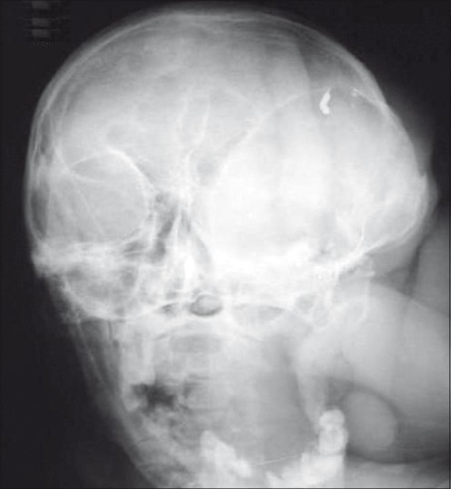
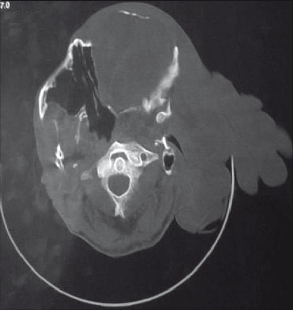
Plain X-ray of the skull showed sphenoid dysplasia [Figure 2]. Dynamic X-rays of the cervical spine showed reducible atlantoaxial dislocation (AAD). Computed tomography (CT) scan of the craniovertebral junction (CVJ) revealed AAD and rotational subluxation [Figure 3].

- Plain X-ray of the skull showed sphenoid dysplasia

- CT scan of the CVJ revealed AAD
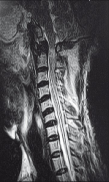
Magnetic resonance imaging of the cervical spine showed AAD, cord compression at CVJ and syrinx from C2 to C7 [Figure 4]. The patient was diagnosed to have NF1 with giant PNF. The atlantoaxial and atlanto occipital junction, though very stable complexes, also have an extensive mobility making them vulnerable for dislocation. Atlantoaxial instability may result from many reasons including hypoplasia of the odontoid process and from laxity of the transverse ligament.[1] In our patient, the progressive enlargement of the PNF with subsequent increase in its weight could have led to the atlantoaxial instability. PNFs are benign tumors originating from nerve sheath cells, subcutaneous, or visceral peripheral nerves, occurring in only 5% of patients with NF1.[2] There are case reports of atlanto axial instability due to neurofibromatosis.[34]

- Magnetic resonance imaging of the cervical spine showed AAD, cord compression at CVJ and syrinx from C2 to C7
The patient presented at admission a Nurich's grade 3 neurological condition. A team of doctors comprising neurologists, neurosurgeon, plastic surgeon and surgical oncology counseled the patient about his condition and the patient opted for the spinal surgery. The patient underwent operation with occiput to C2 and C3 fusion (titanium loop and bone graft) and C1 arch excision. We present this case to highlight a rare manifestation of massive plexiform neurofibroma causing atlantoaxial instability leading to spastic quadriparesis.
References
- Anatomy and physiology of congenital spinal lesions. In: Benzel EC, ed. Spine surgery: Techniques, complication avoidance, and management (2nd ed). Amsterdam: Elsevier; 2005. p. :61-87.
- [Google Scholar]
- The neurofibromatoses. In: Freedberg IM, Eisen AZ, Wolff K, Austen KF, Goldsmith LA, Kartz SI, eds. Fitzpatrick's Dermatology in General Medicine. New York: Mc graw Hill; 1999. p. :2152-8.
- [Google Scholar]
- Atlanto axial instability due to neurofibromatosis: Case report. Acta Orthop Belg. 2000;66:392-6.
- [Google Scholar]
- Atlantoaxial dislocation associated with neurofibromatosis. Report of three cases. J Neurosurg. 1983;58:451-3.
- [Google Scholar]


