Translate this page into:
Epidural hematoma with detachment of the dural sinuses
Address for correspondence: Dr. Federico Caporlingua, Via Gennaro Cassiani 10, Roma – 00155, Italy. E-mail: capor51@gmail.com
This is an open-access article distributed under the terms of the Creative Commons Attribution-Noncommercial-Share Alike 3.0 Unported, which permits unrestricted use, distribution, and reproduction in any medium, provided the original work is properly cited.
This article was originally published by Medknow Publications & Media Pvt Ltd and was migrated to Scientific Scholar after the change of Publisher.
Abstract
Epidural hematoma (EH) is a neurosurgical emergency that requires early surgical treatment. It is rarely extended bilaterally causing a detachment of the dural sinus or sinuses. The authors present two rare cases of EH with dural sinus detachment and describe how they suspend them. In these cases it is crucial to firmly suspend the dura mater and the dural sinus to the inner skull surface to prevent postoperative rebleeding.
Keywords
Extradural hematoma
head injuries
operative technique
Introduction
Extradural hematoma (EH) represents 1% of head trauma admissions. It is more frequent in male population,[1] and usually occurs in the younger population probably because the dura mater of elder patients adheres more to the inner surface of the skull. Nearly 85% of the cases are due to skull fracture with rupture of the middle meningeal artery or its branches.[2]
EH presents as a bilateral hemorrhage in 2%-10% of the cases.[134] In these cases it may be possible to find either two different or one single fracture, which might extend to the contralateral side. However, the hemorrhages do not involve the midline. The first such case was reported by Roy in 1884.[5]
EH may be located in the midline, thus being associated with a detachment of the sagittal longitudinal sinus (SLS).[67] The international literature contains a certain number of EH cases of nonarterial origin. In these cases, the bleeding can originate from rupture of arachnoid granulations, diploic emissary veins, and dural sinuses. They have been described as bifrontal as they detach the first third of the sagittal sinus, biparietal or vertex EH,[267] and bioccipital as they detach the posterior third of the SLS.[8]
Although it is a rare occurrence, either a bioccipital EH or a posterior fossa EH may detach the torcular herophili and/or the transverse sinus.[891011]
The peculiarity of the detachment of the dural sinuses adds a new issue for the neurosurgeon who must suspend the sinus to the inner surface of the skull in order to prevent postoperative rebleeding. We therefore describe an original technical strategy.
Patients and Methods
The authors commonly operate on patients affected by posttraumatic EH. Two cases with involvement of the dural sinuses are described below: One with involvement of the anterior third of the SLS and the other with detachment of the posterior third of the SLS, of the transverse sinuses with consequent extension in the posterior fossa.
Illustrative cases
Case 1
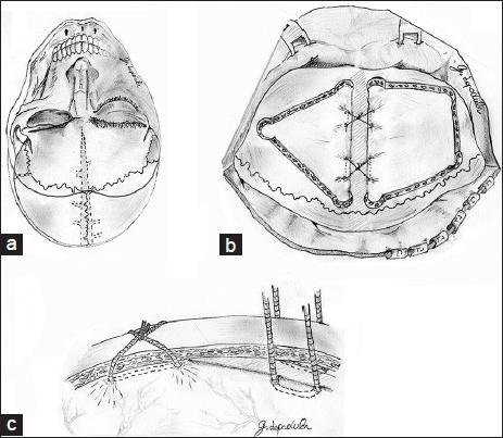
A 23-year-old woman presented in the E.R.emergency department with Glasgow Coma Scale (GCS) 12. A brain computed tomography (CT) scan showed a large frontotemporal right EH with median extension, detachment of the SLS [Figure 1], and a frontotemporoparietal closed fracture. A bicoronal skin flap [Figure 2a] with exposition of the whole frontal region was performed. Four burr holes were drilled on the right side of which two were paramedian to the SLS. The burr holes were drilled with the high-speed drill bur rather than the perforator to prevent pressure on the fractured segment. Through this first craniotomy, most of the hematoma was removed and a few distal arterial lacerations were repaired. Then, a second craniotomy was performed by means of three burr holes on the contralateral side leaving a 3-cm strip bone over the SLS [Figure 2b]. The hematoma was completely removed and the bleeding was stopped with packing and with a hemostatic agent. To suspend the detached dura and sinus, two dural hitch stiches were passed on each side along a parasagittal line to the sagittal sinus, on both sides. The dural stiches were gently raised toward the skull; the SLS was positioned back into its subperiosteal loggia tying the stitches onto the external surface of the bone strip [Figure 2c]. Postoperative course was uneventful and the patients was discharged 4 days after the intervention with no neurological deficit.

- A computed tomography scan showing a right frontal extradural hematoma with contralateral extension and detachment of the sagittal longitudinal sinus

- Illustration (a) of the patient position according to surgeon's view; a bicoronal skin incision flap is traced. Surgical exposure (b) and illustration of the craniotomies after the blood clot evacuation and hemostasis; only the suspensions for the dural sinus are drawn. The dural suspensions (c) are being tied above the strip of bone
Case 2
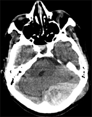
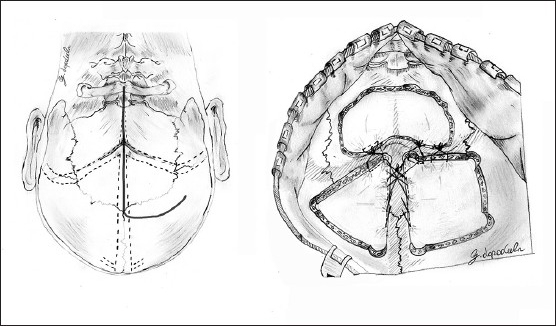
A 56-year-old man presented to our observation in a state of coma after a head trauma with occipital fracture. GCS was 8. A brain CT scan documented a left posterior fossa extradural hematoma with detachment of the left transverse sinus, of the sinus confluence and of the posterior third of the sagittal sinus with extension in the right occipital region [Figure 3]. The hematoma compressed the structures of the posterior cranial fossa and caused a herniation of the fossa content through the foramen magnum and the incisura tentoria. Thus, by means of a right “hockey stick” skin incision flap [Figure 4, left], one suboccipital craniotomy was performed in order to rapidly decompress the posterior cranial fossa and carry out an initial hemostasis. Then, two parieto-occipital craniotomies were drilled; one square-shaped with parasagittal burr holes positioned to the right of the posterior third of the sagittal sinus, and one, triangle-shaped, to the contralateral side leaving a 3-cm strip of bone above the sagittal sinus and the transverse sinuses [Figure 4, right]. Four dural hitch stitches were placed along a paramedian line, on each side of the SLS and of the right transverse sinus and two dural stitches were placed paramedian to the left transverse sinus. The dural stiches were gently raised toward the inner skull plane and secured onto the bone strip that was left above the sinuses [Figure 2c]. Subdural exploration was carried through a small dural opening on the posterior cranial fossa to exclude any subdural hemorrhage. Postoperative course was uneventful.

- A computed tomography scan showing a left cerebellar extradural hematoma. The hematoma extended to the supratentorial space and detached the sinus confluence, the left transverse sinus, and posterior third of the sagittal longitudinal sinus

- Illustration of the patient position according to surgeon's view, a “hockey stick” skin incision flap is traced (left). Surgical exposure and illustration of the suboccipital craniectomy and of the supratentorial craniotomies after the blood clot evacuation and hemostasis (right). In the sketch, only the suspensions for the dural sinus are drawn
Discussion
A classic localization of an EH is temporal with involvement of Marchant's area, where the middle meningeal artery has a bigger diameter and the pressure from the ruptured artery can easily detach the dura mater. The more the EH is located toward the midline, the slower the bleeding may be and, once surgically exposed, the external surface of the dura mater may present with diffuse bleeding. Often it is not possible to recognize a specific bleeding spot. In every case the surgical goal is to decompress the neural structures and to arrest the bleeding. If the middle meningeal artery is ruptured in a proximal site toward the foramen spinosus, it can be exposed and clipped. If multiple and distal bleeding spots are exposed, they can be electrocoagulated, clipped, and stopped with the aid of hemostatic agents. In every case the suspension of the dura mater is essential, where it guarantees a mechanical tamponade, thus minimizing the risk of postoperative rebleeding.
To date, the literature contains a certain number of EH with detachment of the dural sinuses.[12610111213] Yilmazlar et al.[12] described a frequency of 25% of nonarterial EH in their series of 30 patients, among whom 60% were caused by rupture of dural sinuses. Four patients with dural sinus tear were reoperated on because of recurrent hemorrhage. They report a higher recurrence of bleeding in patients harboring dural sinus hematomas and propose ligation of the anterior third of the SLS if necessary. It is common practice to expose the sinus in order to facilitate the use of hemostatic agents and eventually repair the lacerations.
Other surgical techniques that have been described are the direct stitching according to Krause, the free duraplasty, hitching up the dura to the bone adjacent to the sinus, clipping of the sinus, the pedunculated duraplasty.[14] Our technique has the advantages of being easier, faster, and allows to maintain a rigid structure (the bony bridge upon the sinus) to facilitate the brain expansion and to tamponade through suspension the dural tear.
Supratentorial midline EH
When dealing with a bifrontal EH, a coronal skin flap is prepared. A frontal craniotomy is drilled by means of four burr holes. The craniotomy would be located on the side where the hematoma is more represented, would not be extended to the contralateral side and would permit to control most of the bleeding. Successively, a contralateral craniotomy by means of three burr holes is drilled leaving a strip of bone of a few centimeters on the detached dural sinus. The craniotomies permit to achieve a complete control of the bleeding. The dural hitch stiches are passed bilaterally along the SLS. They are not passed through the outer layer of the cortical bone as it has been previously described by Jones et al.[7] as the authors of the present study think that this could cause an excessive and asymmetrical tension on the SLS during its suspension. The SLS is suspended tying the dural stiches onto the strip surface, as shown in Figure 2c.
As for vertex and bioccipital EH, the authors of the present study would use the same technical strategy. However, in case of bioccipital EH, the skin flap would be prepared with respect to the vasculature.
Supra- and subtentorial EH with detachment of the transverse sinus and/or of the torcular herophili
To the authors’ knowledge, 10 cases of supra-and subtentorial EH with detachment of the transverse sinus and\or of the turcular herophili have been reported.[891011] Jamieson et al.[1] reported three cases of “vertical extradural hematoma” in their series, but they do not specify if the EH involved the supra- and the subtentorial space. The authors, in such cases, drill a suboccipital craniectomy and two supratentorial craniotomies on each side of the SLS. The surgical exposure provided by a “hockey stick” incision flap permits to drill a larger craniotomy to the side where the EH is more represented and a smaller one to the contralateral side. If the hematoma detaches only the transverse sinus, being monolateral, two craniotomies on both sides of the transverse sinus will be sufficient. In every case, a 3-cm strip of bone is left as a plan of suspension for the dural sinuses as described before. The dural stitches are passed on each side of the strip of bone and then tied on its outer face, as showed in the second illustrative case.
Conclusion
The authors believe that leaving a strip of bone above the dural sinus aids to create a plane of suspension for the detached dura. This surgical technique is safer, especially during the suspension of the dural sinus, more feasible, and less time-consuming.
Acknowledgment
Gennaro Lapadula and Federico Caporlingua put the same effort in the draft of the present manuscript, thus they are to be considered coauthors.
Source of Support: Nil.
Conflict of Interest: None declared.
References
- Extradural hematoma: Observation in 125 cases. 1960. Lancet. 276:167-72. http://dx.doi.org/10.1016/S0140-6736(60) 91322-2
- [Google Scholar]
- Fracture of skull, extensive extravasation of blood on dura mater, producing compression of brain, trepanning, partial relief of symptoms, death. Lancet. 1884;2:319.
- [Google Scholar]
- A surgical strategy for vertex epidural haematoma. Acta Neurochir (Wien). 2011;153:1819-20.
- [Google Scholar]
- Bilateral extradural hematoma extending from the foramen magnum to the vertex. Surg Neurol. 1987;28:123-8.
- [Google Scholar]
- Bilateral posterior fossa epidural hematoma-report of two cases. Neurol Med Chir (Tokyo). 1992;32:690-2.
- [Google Scholar]
- Study on cases with posterior fossa epidural hematoma - Clinical features and indications for operation. Neurol Med Chir (Tokyo). 1990;30:24-8.
- [Google Scholar]
- Traumatic epidural haematomas of nonarterial origin: Analysis of 30 consecutive cases. Acta Neurchir (Wien). 2005;147:1241-8.
- [Google Scholar]
- Bilateral frontal extradural haematomas caused by rupture of the superior sagittal sinus: Case report. Br J Neurosurg. 1999;13:77-8.
- [Google Scholar]
- The traumatic dural sinus injury-a clinical study. Acta Neurochir (Wien). 1992;119:91-3.
- [Google Scholar]


