Translate this page into:
Advanced documentation methods: Putting a new spin on the traditional wheel
*Corresponding author: Dr. Archana Sharma, Department of Neuroanesthesia and Neurocritical Care, National Institute of Mental Health and Neurosciences, Bengaluru, Karnataka, India. drarchana7anesthesia@gmail.com
-
Received: ,
Accepted: ,
How to cite this article: Sharma A, Chakrabarti D, Surve R, Gandhi M. Advanced documentation methods: Putting a new spin on the traditional wheel. J Neurosci Rural Pract. 2025;16:93-5. doi: 10.25259/JNRP_295_2024
Abstract
Documenting the intraoperative proceedings is an important yet relatively relentless task for practicing anesthesiologists. This paper reports a novel method to document the monitoring data in peri-operative periods and critical care settings that will help clinicians record and retrieve monitoring data for research and medicolegal purposes. This novel technique is inexpensive, effective, can be used in various settings (urban or rural practice), and will help clinicians in every cadre.
Keywords
Advanced documentation
Anesthesia research
Data retrieval
Perioperative monitoring
INTRODUCTION
Documentation of intraoperative proceedings is an important yet relatively arduous task for practicing anesthesiologists. The data is essential for medicolegal purposes and for conducting research efficiently (especially retrospectively). High-frequency recording of intraoperative monitoring parameters is impossible to conduct manually. Low-frequency data cannot tease the causation behind intraoperative adverse events and is insufficient for research. For example, venous air embolism, detected by a sudden drop in end-tidal CO2, occurs within seconds, where even a 1-minute data capture window would lose out on figuring out the timeline of events. High-frequency data capture methods currently involve high equipment, software, and maintenance costs. Since such methods are technology-heavy, simple requirements like data extraction for research are difficult for the average clinician. These software are customized to the equipment used in the operation theaters. Any change in equipment requires a high cost of upgradation. This paper highlights a simple, effective, low-cost technique to acquire high-frequency data for archival and research purposes.
Every monitor is a computer with a processing component and a screen displaying the processed data. This generalization can be leveraged to suit our needs by routing the processed data to a screen of our choice, wherein the output can be archived easily. Utilizing this principle, we describe a methodology tested on multiple monitor systems of various configurations to effectively stream and save the monitoring data to a Microsoft Windows operating system platform.
The required hardware includes:
Windows computer - at least 4 GB random-access memory, 4 GB graphics card (equivalent to NVIDIA GT735 or higher), and a mid-range processor (equivalent to IntelTM i5-3400 or higher) with USB 3.0 or higher port.
A video capture card capable of 30 frames/second capture at 1080 p (1920 × 1080 pixel resolution).
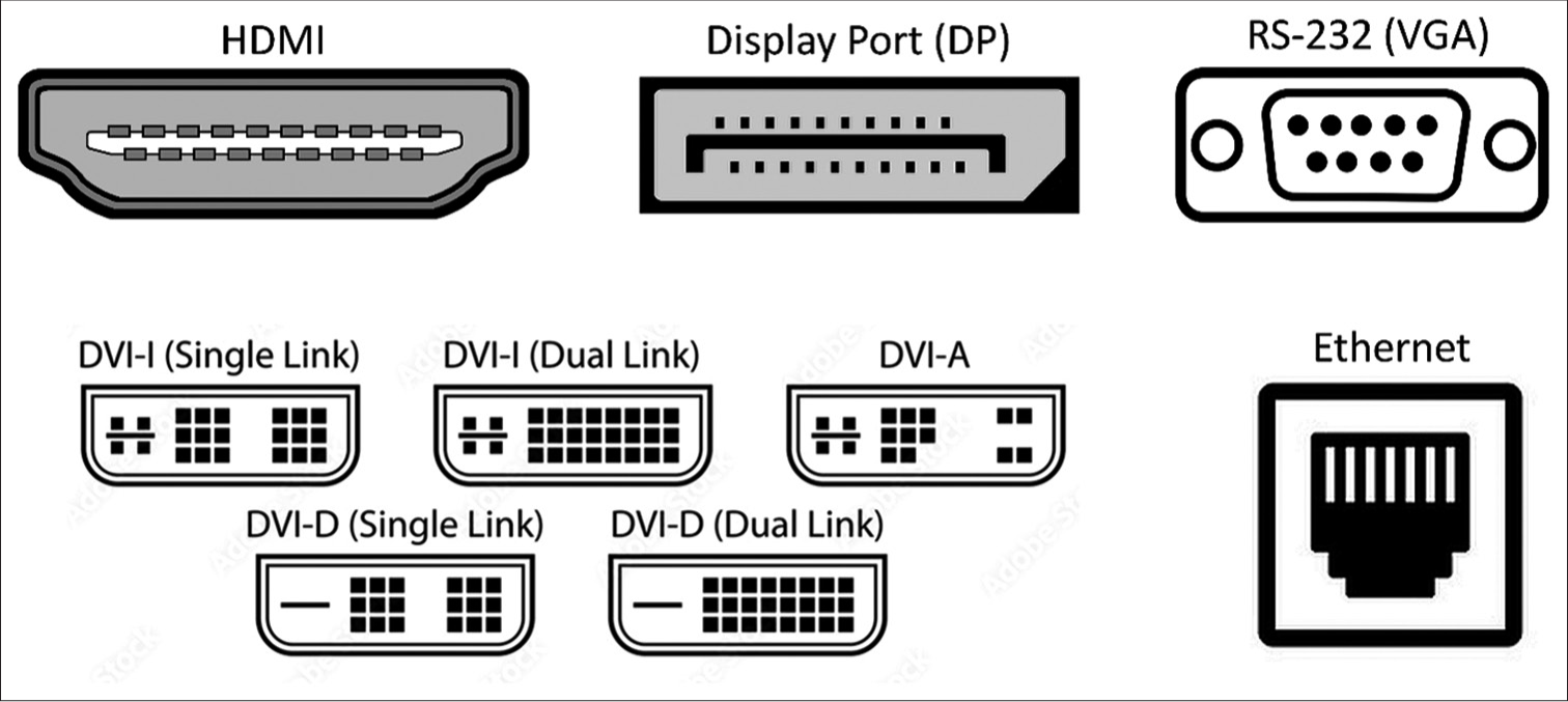
Adaptors and cables for monitor-specific video output ports to High-Definition Multimedia Interface (HDMI) (usual input port in video capture cards) [Figure 1].

- Vector images representing common types of video-out ports in multiparameter monitors.
The software required is the open-source VideoLAN Client (VLC) media player by the VideoLAN organization.[1]
STEPS TO BE FOLLOWED
Configure VLC media player: Press Ctrl + P to open “Preferences” [Flowchart and Video]. In the left lower corner, “Show settings,” click “All.” In the left scrolling panel, click “Input/Codecs” -> “Video codecs” -> “FFmpeg.” In the right panel, “Hardware decoding,” click “Disable” in the drop-down menu. Below that, increase “Threads” to 2. Save preferences by clicking “Save.” Close and reopen VLC (these changes only need to be done once, in the beginning). Find the type of video out port in the multiparameter monitor. Usually, it is either HDMI, display port (DP), Digital Visual Interface, RS-232, or Ethernet port [Figure 1]. The type of adapter required will be based on this port. These adaptors are relatively inexpensive. Connect the adaptor and cable to the monitor port at the one end and the video capture card HDMI port at the other. Connect the video capture card to your computer through USB.
On VLC, press Ctrl + C to open the “Open Media” widget. For “Video device name,” open the drop-down menu and find and select the option for your capture card – “USB3. 0 capture.” Under video size, type the pixel dimensions of the monitor to be captured in “height-width” format. For example, “1280 × 720” or “640 × 480”. Leaving it blank will default to “640 × 480”. Click “Advanced options” and modify the “Picture aspect ratio” if required. Usual configurations are “4:3” or “16:9”. This will change the final output video’s aspect ratio (height: width). The output video will be coerced into the set aspect ratio. For “video input frame rate,” leaving at 0 will allow the capture at a frame rate that best suits your system. For high-quality video, the rates supported by inexpensive capture cards are capped at 30 frames/second. Higher frame rates entail higher storage space requirements. All other options in the “Advanced options” box must be left as they are. On the “Open Media” screen, if only visualization of the monitor screen is required, press “Play” and the streaming should start.
If recording of the screen is required, click the drop-down menu adjacent to “Play” and click “Convert.” On the “Convert” screen, click “Display the output.” Click the spanner button next to “Profile.” This opens the “Profile edition” window; under the “Encapsulation” tab, click “MP4/MOV.” Under the “Video codec” tab, click the “Video” option and press the “Save” tab at the bottom. Back on the “Convert” screen, click the “Browse” button in front of “Destination file:” and choose the directory in which to save the video file and provide a name for the video file. Upon clicking “Start,” the video streaming and saving will begin. If a blank screen appears while the timer at the bottom is running, the video is being saved. However, the computer does not have enough computing resources to display and save simultaneously. The video recording can be stopped by clicking the “square” stop button at the bottom of the VLC screen.
DISCUSSION
Incentive compensation management (ICM) software[2] was developed in 1986, for collecting data from bedside monitors to describe cerebral autoregulation and pressure-volume compensation; it has now been extended to ICM + which records raw signals and calculates time trends of summary parameters, displayed in simple time trends, as well as time window-based histograms, cross histograms, correlations, etc. All this allows complex information coming off the bedside monitors to be summarized and presented to medical and nursing staff in a simple way that alerts them to the development of various pathological processes.
We simplified the setup and procedure to cut down the cost and time to an insignificant level. The uses of this method are limited by one’s imagination. It can be implemented at a very low cost and has wide applicability across multiple monitor systems. For an anticipated difficult case, the monitor screen can be recorded for the procedure’s duration to archive the smallest changes in the patient’s vitals. It can also aid in substantiating evidence in medicolegal proceedings or for teaching purposes. Suppose a research project requires recording monitor parameters at a higher frequency. In that case, the video recording will ensure high-quality data and numeric values retrieval later at one’s leisure, devoid of reflected light glare, or contrast issues observed in images of the screen clicked by a camera. The authors of this article are also working on automated methods of recording high-frequency numeric data from video recordings of intraoperative screens and converting it into an Automated Excel sheet. The authors believe that another software Open Broadcaster Software Studio[3] is a free and open-source screen casting and streaming application that is compatible with both Windows and macOS and can be utilized as a substitute for a VLC media player.
The series of steps described may take a little practice to get used to, but once refined for one’s setup, it takes very little time to start quickly. Embracing and adapting readily available and open-source technology can go far in inexpensively establishing high-quality processes.
CONCLUSION
A simple, effective, low-cost novel technique to acquire high-frequency data for archival and research purposes has been described above that can be used by clinicians of every cadre in urban as well as rural settings.
Ethical approval
Institutional Review Board approval is not required.
Declaration of patient consent
Patient’s consent is not required as there are no patients in this study.
Conflicts of interest
There are no conflicts of interest.
Use of artificial intelligence (AI)-assisted technology for manuscript preparation
The authors confirm that there was no use of artificial intelligence (AI)-assisted technology for assisting in the writing or editing of the manuscript, and no images were manipulated using AI.
Financial support and sponsorship: Nil.
References
- VLC media player. 2006. Available from: https://www.videolan.org/vlc/index.html [Last accessed on 2024 Jul 15]
- [Google Scholar]
- ICM+: Software for on-line analysis of bedside monitoring data after severe head trauma. Acta Neurochir Suppl. 2005;95:43-9.
- [CrossRef] [PubMed] [Google Scholar]
- 2022. Available from: https://obsproject.com/download [Last accessed on 2024 Jul 15]


