
Translate this page into:
A Rare Case of Complete Absence of Mammillary Bodies on Imaging

Nishtha Yadav, MBBS, MD, DM Department of Neuroradiology, Super Speciality Hospital, Netaji Subhash Chandra Bose Medical College Jabalpur 482003, Madhya Pradesh India nishthayadav@yahoo.com
This article was originally published by Thieme Medical and Scientific Publishers Pvt. Ltd. and was migrated to Scientific Scholar after the change of Publisher.
Mammillary bodies act as a relay between hippocampus and upstream structures and have an independent role in memory and contribute particularly to episodic memory.1 Their absence is extremely rare and has previously been described in very few cases.1 2 We report a case with magnetic resonance imaging (MRI) findings of mammillary bodies' absence in a child who underwent imaging for evaluation of seizures.
A 6-year-old male child of healthy nonconguineous parents presented with recurrent episodes of unprovoked falls for 1 month. There was no history of maternal drug intake, irradiation/abnormal event during pregnancy. Birth history was normal with full-term normal vaginal delivery without any history of perinatal insult/birth asphyxia. There was no history of any poor scholastic performance. Routine blood investigations and cerebrospinal fluid (CSF) analysis did not reveal any abnormalities. Interictal electroencephalogram (EEG) revealed periodically generalized epileptiform activity.
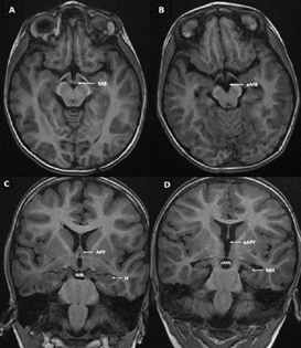
MRI brain revealed complete bilateral absence of mammillary bodies on three-dimensional (3D) T1 images (Fig. 1). Bilateral malrotated vertically oriented hippocampi were noted. There was absence of anterior pillars of fornix.
-
Fig. 1 Axial T1 3D SPGR image showing mammillary body (MB) in control (A), and absence of MB (aMB) in our case (B). Coronal T1 3D SPGR image perpendicular to hippocampus in control (C) showing MB, anterior pillar of fornix (APF) and hippocampus (H) in control. Coronal T1 3D SPGR images in our case (D) shows aMB, aAPF, and malrotated hippocampus (MH). 3D SPGR, three-dimensional spoiled gradient recalled.
Fig. 1 Axial T1 3D SPGR image showing mammillary body (MB) in control (A), and absence of MB (aMB) in our case (B). Coronal T1 3D SPGR image perpendicular to hippocampus in control (C) showing MB, anterior pillar of fornix (APF) and hippocampus (H) in control. Coronal T1 3D SPGR images in our case (D) shows aMB, aAPF, and malrotated hippocampus (MH). 3D SPGR, three-dimensional spoiled gradient recalled.
Detailed cognitive analysis could not be performed as the child was not cooperative.
Complete absence (rather than atrophy) of mammillary bodies is extremely rare. Clinically these patients have developmental amnesia characterized by episodic memory loss.3 To the best of our knowledge, only two cases have been described till date with complete absence of mammillary bodies.1 2 These cases include a patient having developmental amnesia who also had abnormally oriented hippocampi and absence of anterior pillars of fornix.1 Another described case of absent mammillary bodies was associated with the Ohtahara Syndrome (early infantile epileptic encephalopathy with suppression burst) with presence of abnormal orientation of the hippocampi (also noted in our patient) and olivary-dentate dysplasia.2 Both these cases had history of perinatal insult, whereas our patient did not have history of perinatal insult.
MRI findings in our patient completely resemble the case described by Rosenbaum et al who postulated that absence of mammillary body may be a congenital/prenatal developmental abnormality rather than due to perinatal insult.1 The fact that our patient did not have any history of perinatal insult further supports this hypothesis. Additional malformations like vertically oriented hippocampi and absence of pillars of fornix also point toward a developmental malformation.
Other causes of mammillary body atrophy include perinatal insult, thiamine deficiency, and iatrogenic causes (such as after severing of fornix during surgery).3 4 5 6 However, these cases do not have associated symmetric hippocampal and forniceal abnormalities (as noted in our case).
Due to its rarity, little is known about direct behavioral consequences of mammillary bodies' absence. Further detailed neuropsychological evaluation of these patients may provide an insight to the complex memory pathways. Our case adds to the extremely limited literature of congenital absence of mammillary body with hippocampal malformation and forniceal anterior pillar absence. While most cases of mammillary bodies absence are due to acquired causes, however, our case supports the hypothesis that absence of mammillary bodies may also be due to congenital/prenatal abnormality, at least in some cases.
Conflict of Interest
None declared.
Funding None.
References
- Congenital absence of the mammillary bodies: a novel finding in a well-studied case of developmental amnesia. Neuropsychologia. 2014;65:82-87.
- [Google Scholar]
- A case of Ohtahara syndrome with olivary-dentate dysplasia and agenesis of mamillary bodies. Epilepsia. 2001;42(7):950-953.
- [Google Scholar]
- Hippocampal and diencephalic pathology in developmental amnesia. Cortex. 2017;86:33-44.
- [Google Scholar]
- Signal change in the mammillary bodies after perinatal asphyxia. AJNR Am J Neuroradiol. 2019;40(11):1829-1834.
- [Google Scholar]
- Neuroimaging findings in pediatric Wernicke encephalopathy: a review. Neuroradiology. 2010;52(6):523-529.
- [Google Scholar]
- The frequency and extent of mammillary body atrophy associated with surgical removal of a colloid cyst. AJNR Am J Neuroradiol. 2009;30(4):736-743.
- [Google Scholar]


