Translate this page into:
Gun shot injury as a cause of elevated skull fracture
Address for correspondence: Dr. Augustine A Adeolu, Department of Neurological Surgery, Institute of Neurosciences, University College Hospital, Ibadan, Nigeria. E-mail: adeoluaa@yahoo.com
This is an open-access article distributed under the terms of the Creative Commons Attribution-Noncommercial-Share Alike 3.0 Unported, which permits unrestricted use, distribution, and reproduction in any medium, provided the original work is properly cited.
This article was originally published by Medknow Publications & Media Pvt Ltd and was migrated to Scientific Scholar after the change of Publisher.
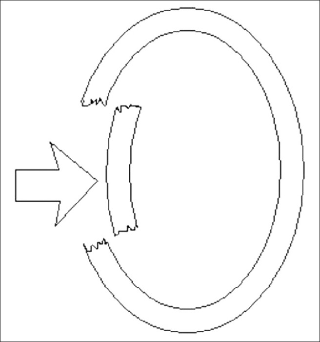
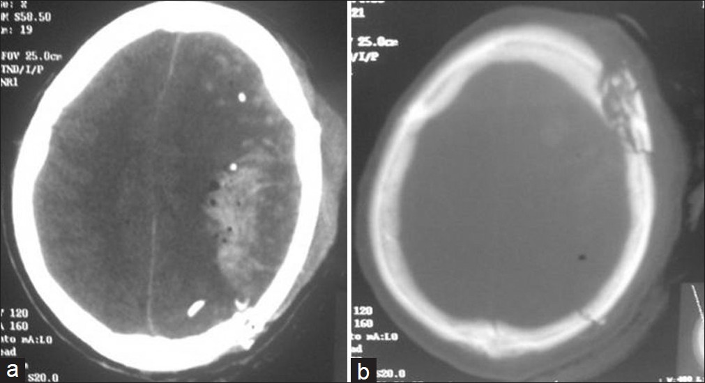
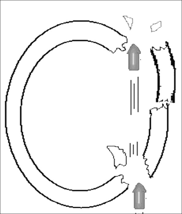
Agents of wounding in head injury often cause displacement of free bone fragment towards the intracranial cavity because of the inward direction of such wounding force. This type of injury is the more common type when a free segment of skull fracture occurs; resulting in what is called depressed skull fracture [Figure 1]. In some rare instances, the free bone fragment is elevated above the intact skull bone. Only few of such cases have been reported in modern literature.[1–5] The common mechanism in these cases was a lateral pull on the free bone fragment arising from a long, thin, relatively sharp and strong object. The mechanism was not certain in other cases, which occurred following road traffic accidents.[13] The following case illustrates another possible pathomechanism of this rare injury. A 39-year-old male truck driver presented to us with loss of consciousness of 4 hour- duration, following a gunshot injury to the head. The shooting range was about 4 meters. His physical examination revealed a young patient with Glasgow coma score of 9 with right faciopariesis and a spastic right hemipariesis. There was diffuse left fronto-parieto-temporal scalp swelling with a left posterior parietal puncture wound and an exit wound at the left frontal region, measuring about 1.5 cm in diameter. Both wounds were discharging altered blood with contused brain matter. The diagnosis was gunshot injury to the head with moderate head injury and bihemispheric deficits, worse in the left hemisphere. The cranial computerized tomography scan showed extensive left hemispheric multifocal hemorrhagic contusions, multiple intra-axial bony fragments and diffuse brain swelling with midline shift to the right. There was a left posterior parietal skull perforation with communited, elevated left frontal fracture. [Figure 2] A left fronto-parieto-temporal decompressive craniectomy and wound debridement was performed. The findings at surgery were: An elevated left frontal and linear left fronto-parietal skull fractures as well as left hemispheric cortical contusions. During the post-operative period, global aphasia and dense right spastic hemipariesis were noted, and these resolved gradually. The patient has made progressive recovery with resolution of his aphasia, and he currently walks with no support. Our patient's injury was the perforating gunshot type, in contrast to the elongated sharp or blunt weapons that were predominant in earlier case reports.[1–3] The elevated fracture in this case was most likely from the outward force, impacted on the frontal bone from the bullet that was exiting the intracranial cavity. This is further illustrated in Figure 3. Elevated skull fractures have been proven to be a distinct entity with both imaging modalities and surgical findings. It often results from a significant centrifugal force applied to the head with a weapon, often causing significant structural injury, neurologic deficits and sequela. Most cases are compound fracture, requiring surgical intervention.

- Diagrammatic representation of depressed skull fracture. The arrow represents the applied force causing the fracture with displacement of the floating segment inwards

- Cranial CT scan, brain and bone windows

- Elevated skull fracture from gunshot head injury
Source of Support: Nil
Conflict of Interest: None declared.
References
- Compound elevated fractures of the skull.Report of two cases. J Neurosurg. 1976;44:77-8.
- [Google Scholar]
- Compound elevated skull fracture: A forgotten type of skull fracture. Surg Neurol. 2006;65:503-5.
- [Google Scholar]
- Elevated skull fractures in pediatric age group: Report of two cases. Turk Neurosurg. 2011;2:418-20.
- [Google Scholar]
- Post- traumatic compound elevated fracture of skull simulating a formal craniotomy. Turk Neurosurg. 2009;19:103-5.
- [Google Scholar]


