Translate this page into:
Computerized tomographic study of normal Evans index in adult Nigerians
Address for correspondence: Dr. SA Olarinoye-Akorede, Department of Radiology, Ahmadu Bello University Teaching Hospital, Zaria, Kaduna, Nigeria. E-mail: olarinoyebs@yahoo.com
This is an open-access article distributed under the terms of the Creative Commons Attribution-Noncommercial-Share Alike 3.0 Unported, which permits unrestricted use, distribution, and reproduction in any medium, provided the original work is properly cited.
This article was originally published by Medknow Publications & Media Pvt Ltd and was migrated to Scientific Scholar after the change of Publisher.
Abstract
Background:
The evaluation of degree of ventricular enlargement should be based on established indices rather than on personal experience as this is highly subjective. Our aim was to establish normal values for Evans index in a Nigerian adult population as none has been found in the Nigerian medical literature.
Materials and Methods:
Axial computerized tomographic brain scans of 488 normal subjects were reviewed retrospectively. Of them, 319 (65.36%) of the patients were males and 169 (34.63%) were females; their ages ranged from 18 to 84 years with a mean age of 37.26 years. The images were acquired using a multi-slice GE Sigma excite scanner. Evans index was measured as the linear ratio of the total width of the frontal horns of the cerebral lateral ventricles to the maximum intracranial diameter.
Results:
The mean value for Evans index for the studied population was 0.252 ± 0.04. The EI increased with age and it was slightly higher among males. The difference in Evans value in males and females was not statistically significant. Individuals above 60 years old had the highest Evans values in both sexes.
Conclusion:
This study has established ranges of normal value for Evans index in a Nigerian population. It agrees with the diagnostic cut-off value of > 0.3 for hydrocephalus and it compares well with that of the Caucasians.
Keywords
Computerized tomography
Evans index
Nigeria
normal
normal adults
Introduction
Ventricular enlargement is a characteristic physical change that is present frequently in a number of cerebral disorders encountered in neurologic and psychiatric practice. It is probably the most common encephalographic abnormality in both children and adults.[1] Often times, the assessment of ventricular enlargement when viewing images from computerized tomography (CT) or magnetic resonance imaging (MRI) is based on the subjective assessment of the reviewer's experience. Older radiographic methods (gas or contrast encephalography) were once employed but these were invasive and also introduced artifacts.[2] Some of the limitations have been overcome by the newer neuroimaging techniques like CT and MRI and this has led to better attempts at evaluating ventricular sizes.[345] Evans index (EI) is one of the ventriculographic indices used. It is a ratio which compares the maximum width of the frontal horns of the lateral ventricle to the maximum transverse diameter of the inner table of the skull. It also serves as an indirect marker of ventricular volume.[67] EI has been extensively used in the diagnosis of idiopathic normal pressure hydrocephalus, in the assessment of outcome of patients with shunt placement which is the primary mode of treatment[4] and also in the assessment of visual complications of childhood hydrocephalus.[8]
Materials and Methods
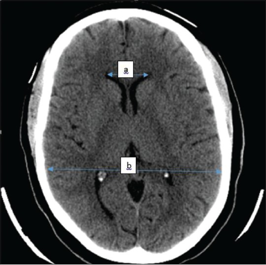
Ethical consideration does not permit that healthy individuals with no clinical symptoms be subjected to ionizing radiation. Therefore, CT scans of neurologic patients which were reported to be normal were reviewed for this study. Being a retrospective study, patient consent was not required however; this research was approved by the ethics committee of Ahmadu Bello University Teaching Hospital. Scan reports of 492 patients that were reported as normal were reviewed independently alongside their images by two radiologists. However, a total of 488 patients were finally selected and were analyzed for the study. They consisted of 319 males and 169 females and ranged between 18 and 84 years old. CT images were obtained from the local database of the CT machine and back up compact discs from the CT archives. All patients had two-phase examination (pre and post contrast studies) of the brain using the department's Siemens CT scanner (Sigma Excite HD) at 2.5 mm slice thickness at the skull base then 5 mm to the vertex. The images were viewed on the computer monitor using a meter rule with which the following measurements were made as seen in Figure 1.

- Illustrative axial CT section of one of the selected patients showing how measurements were taken Total anterior horn width (TAHW) =a Maximum intracranial diameter (MICD) =b
Total Anterior horn width (TAHW): a
Maximum intracranial diameter (MICD): b
Evans index (EI) derived by calculation for each patient as:

Exclusion criteria
Three patients who had only pre-contrast examination were not included in the study population. The absence of positive finding in a single phase examination was considered inconclusive. Also, one patient was not selected who had only descriptive report of normal without a final conclusion.
Statistical analysis
Statistical analysis was done using Sigmastat 2.0 for windows San Rafael CA. Statsoft (1995). Data are presented in tables and charts, and expressed as percentages, means and standard deviation. Differences in ventricular parameters with respect to sex were examined using Student's t test. One-way analysis of variance was used to analyze for variations across age groups. P < 0.05 was considered as statistically significant.
Results
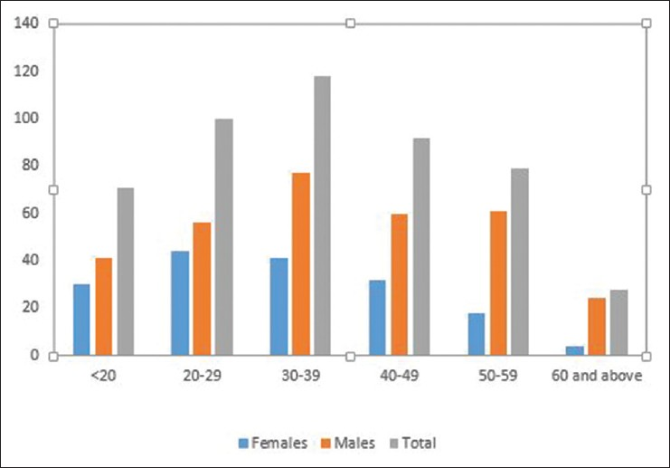
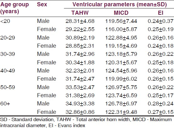
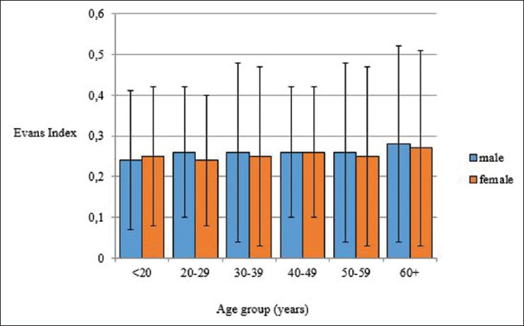
These results were obtained from the analysis of 488 patients that were finally selected; their age and sex distribution is seen in Figure 2 there were more males in this study with a male: female ratio of 1.88:1. Most patients were between 30 and 39 years and their mean age was 37.26 years. The difference in age between males and females was statistically significant (P < 0.001). Table 1 describes some ventricular parameters with respect to sex, while in Table 2 EI, total anterior horn width and maximum intracranial diameters are shown with respect to age and sex of the patients. The distribution of EI among the different age groups in both sexes is depicted in a grouped bar chart in Figure 3, the bars representing the standard deviation. The mean Evans EI in the studied population was 0.257 ± 0.04 in males, 0.256 ± 0.04 in the females and an overall mean of 0.252 ± 0.04. There was no significant statistical difference in the Evans ratio between male and females (P = 0.08). This study shows that EI was highest in individuals who were 60 years and above in both males and females.

- Age and sex distribution of the patients


- Grouped bar chart of Evans index versus age group and sex
Discussion
Ventricular enlargement in adults could be as a result of aging, neurodegenerative diseases, cerebro-vascular conditions, tumors and trauma. In our local literature, description of ventricular sizes are done mostly in children,[89101112] while in adults it is under-reported. EI is a quantitative criterion which has been used extensively in assessing ventriculomegaly and the mean value in this study agrees with those of previous Caucasian studies.[5671314] We also found that EI increased with advancing age as reported by other authors.[1314] This is due to the fact that the brain substance shrinks with age, while the cerebrospinal fluid spaces which include the ventricles increase in size in order to compensate for the atrophying brain parenchyma. With this physiologic ventricular enlargement, the Evans ratio does not exceed 0.3. We did not find a statistically significant difference in Evans ratio between males and females. Haug[15] on the other hand reported a smaller ventricular system in females than in males in individuals above 15 years old while the reverse was the case in individuals below 15 years old in the same study. Scrutinous search of the literature revealed poverty of Nigerian reports on the assessment, diagnosis and prognosis of adult hydrocephalus. The authors advocate a need for its early detection especially in adults and the elderly since cranial enlargement which could have been a pointer is not possible in the fused adult skull. The distressful effects of prolonged severe cerebral atrophy on memory, cognitive ability and autonomic function could then be avoided especially because some of the causes are treatable.
Idiopathic normal pressure hydrocephalus is a potentially reversible cause of dementia in the elderly which responds positively to CSF shunting procedures. The syndrome comprises of ventricular enlargement, gait disturbance and urinary incontinence.[5] Several imaging parameters are considered in order to make a diagnosis which include: Tight high cerebral convexity (best seen on coronal MRI), medial subarachnoid spaces, and enlarged Sylvian fissures.[16] These are subjective and nonspecific.[4] Apart from that, the examiner is also faced with the problem of proving that the degree of hydrocephalus is in excess of the degree of atrophy that is present. EI as measured by CT or MRI on the other hand quantitatively assesses the degree of ventricular enlargement, with an international guideline diagnostic cut off value of >0.3.[7] EI in the presence of the clinical symptoms could be sufficient for the diagnosis. Our finding also supports that this defining criterion (EI > 0.3) could be used in the diagnosis of hydrocephalus in our own environment. Poca et al. found EI to be of good predictive index in post-traumatic ventriculomegaly[17] while Odebode et al. employed it in assessing the relationship of ventricular size with visual function in children with hydrocephalus.[10]
One of the limitations of the isolated use of EI is that it only assesses the degree of hydrocephalus but does not give information about extent of cerebral atrophy.[5] Nonetheless, EI when used with other derived indices e.g., callosal angle,[18] frontal horn index[7] has proved valuable in examining CSF or subarachnoid spaces. It is less technical than other sophisticated methods, less time consuming and can be used in routine practice.
Studies by Ambarki et al.[6] and Toma et al.[7] showed strong correlation of EI with ventricular volume r = 0.94 and r = 0.619, respectively. They however still suggest direct volumetric measurements while Bezing et al. advocate that in patients with hydrocephalus following tuberculous meningitis, linear measurements are more reliable than volumetric ratios.[19] In the elderly, it is even more imperative to differentiate between the various causes of ventricular enlargement which could have overlapping clinical symptoms e.g. normal ageing, Alzheimer's disease, idiopathic normal pressure hydrocephalus, Parkinson's disease and dementia with Lewi bodies.[20] Future studies will be necessary for this distinction because accurate and timely diagnosis is essential for successful treatment. Recent attempts include direct volumetric assessment using sophisticated software algorithms like the mean diffusivity histogram analysis,[20] the use of CSF biomarkers, Positron emission tomography with amyloid tracers, total and phosphorylated Tau measurements.[2122] These techniques are not yet available in our environment and even in the developed countries, it is limited by cost.
Conclusion
In this presentation, the authors have determined the range of normal values of Evans index in a Northern Nigerian population using computerized tomography.
Source of Support: Nil.
Conflict of Interest: None declared.
References
- An encephalographic ratio for estimating ventricular enlargement and cerebral atrophy. Arch Neurol Psychiatry. 1942;47:931-7.
- [Google Scholar]
- Computerized tomography (The EMI scanner): A comparison with pneumoencephalography and ventriculography. J Neurol Neurosurg Psychiatry. 1976;39:203-11.
- [Google Scholar]
- Apparent diffusion coefficient and cerebrospinal fluid flow measurements in patients with hydrocephalus. J Comput Assist Tomogr. 2008;32:392-6.
- [Google Scholar]
- Study of INPH on Neurological Improvement (SINPHONI). Diagnosis of idiopathic Normal pressure hydrocephalus is supported by MRI-based scheme: A prospective cohort study. Cerebrospinal Fluid Res. 2010;7:18.
- [Google Scholar]
- A pilot study of quantitative MRI measurements of ventricular volume and cortical atrophy for the differential diagnosis of normal pressure hydrocephalus. Neurol Res Int. 2012;2012:718150.
- [Google Scholar]
- Brain ventricular size in healthy elderly: Comparison between Evans index and volume measurement. Neurosurgery. 2010;67:94-9.
- [Google Scholar]
- Evans’ index revisited: The need for an alternative in normal pressure hydrocephalus. Neurosurgery. 2011;68:939-44.
- [Google Scholar]
- Visual function in infants with congenital hydrocephalus with and without myelomeningocoele. Childs Nerv Syst. 2014;30:327-30.
- [Google Scholar]
- Etiology and cranial CT scan profile of nontumoral hydrocephalus in a tertiary black African hospital. J Neurosurg Pediatr. 2011;7:397-400.
- [Google Scholar]
- The relationship between ventricular size and visual function in children with hydrocephalus. Afr J Med Med Sci. 1998;27:213-8.
- [Google Scholar]
- Intracranial ventricular sizes and correlates in term Nigerian infantsat birth and six weeks. Afr J Med Med Sci. 1988;27:213-8.
- [Google Scholar]
- Chiari I anatomy after ventriculoperitoneal shunting: Posterior fossa volumetric evaluation with MRI. Childs Nerv Syst. 2006;22:1451-6.
- [Google Scholar]
- Measurements of the normal ventricular system with computer tomography of the brain. A preliminary study on 44 adults. Neuroradiology. 1976;10:205-13.
- [Google Scholar]
- Variations in the size of the human brain. Influence of age, sex, body length, body mass index, alcoholism, Alzheimer changes, and cerebral atherosclerosis. Acta Neurol Scand Suppl. 1985;102:1-94.
- [Google Scholar]
- Age and sex dependence of the size of normal ventricles on computed tomography. Neuroradiology. 1977;14:201-4.
- [Google Scholar]
- Changes in the subarachnoid space precede ventriculomegaly in idiopathic normal pressure hydrocephalus (iNPH) Intern Med. 2012;51:1751-3.
- [Google Scholar]
- Ventricular enlargement after moderate or severe head injury: A frequent and neglected problem. J Neurotrauma. 2005;22:1303-10.
- [Google Scholar]
- Clinical impact of the callosal angle in the diagnosis of idiopathic normal pressure hydrocephalus. Eur Radiol. 2008;18:2678-83.
- [Google Scholar]
- Are linear measurements and computerized volumetric ratios determined from axial MRI useful for diagnosing hydrocephalus in children with tuberculous meningitis? Childs Nerv Syst. 2012;28:79-85.
- [Google Scholar]
- Differential diagnosis of normal pressure hydrocephalus by MRI mean diffusivity histogram analysis. AJNR AmJ Neuroradiol. 2013;34:1168-74.
- [Google Scholar]
- Imaging brain amyloid in Alzheimer's disease with Pittsburgh Compound-B. Ann Neurol. 2004;55:306-19.
- [Google Scholar]
- Alzheimer's Disease Neuroimaging Initiative. Cerebrospinal fluid biomarker signature in Alzheimer's disease neuro imaging initiative subjects. Ann Neurol. 2009;65:403-13.
- [Google Scholar]


