
Translate this page into:
High-voltage electrocution causing bulbar dysfunction
This is an open access article distributed under the terms of the Creative Commons Attribution NonCommercial ShareAlike 3.0 License, which allows others to remix, tweak, and build upon the work non commercially, as long as the author is credited and the new creations are licensed under the identical terms.
This article was originally published by Medknow Publications & Media Pvt Ltd and was migrated to Scientific Scholar after the change of Publisher.
Abstract
Electrical shock can result in neurological complications, involving both peripheral and central nervous systems, which may present immediately or later on. High-voltage electrical injuries are uncommonly reported and may predispose to both immediate and delayed neurologic complications. We report the case of a 68-year-old man who experienced a high-voltage electrocution injury, subsequently developed bulbar dysfunction and spontaneously recovered. We describe the development of bulbar palsy following a significant electrical injury, which showed no evidence of this on magnetic resonance imaging. High-voltage electrocution injuries are a serious problem with potential for both immediate and delayed neurologic sequelae. The existing literature has no reports on bulbar dysfunction following electrocution, apart from motor neuron disease.
Keywords
Bulbar dysfunction
electrocution
high voltage

Introduction
High-voltage electrical injuries are uncommonly reported and may lead to serious neurologic sequelae. The true incidence and prevalence of these events are difficult to ascertain. The extent of injury may be apparent immediately, or it may take weeks to manifest. Clinicians need to be aware of the neurological consequences of electrocution.
Case Report
A 68-year-old male while working on top of building accidentally fell on overhead high-tension cable carrying approximately 11,000 V. He was rescued after a delay of 17 minutes. His burn injury consisted of full thickness burns on the nape of the neck, lower dorsolumbar area [Figure 1]. The patient lost consciousness.
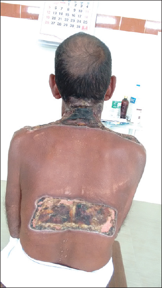
- Full thickness burns injury of the patient over nape of the neck and lower dorsolumbar area
He was admitted in Intensive Care Unit when he was drowsy, minimal response to deep, painful stimulus and was moving limbs to a painful stimulus. He had no signs of cardiovascular or respiratory involvement. His sensorium improved gradually but had occasional disorientation to person, time. His neurological examination revealed no motor or sensory deficit. He was subsequently transferred to surgery ward for the management of burns. Escharotomy was done. A neurological consultation was sent to us on his 15th day of hospitalization because of slurring of speech. On examination, our patient was alert and able to obey commands. A cranial nerve examination revealed reactive pupils, intact extraocular movements, and symmetrical facial movement to smile, eye closure, and eyebrow elevation. However, he had dysarthric speech, dysphonia, and dysphagia. Palatal movements were reduced bilaterally with pooling of secretions and poor gag reflex. There was no associated tongue atrophy or fasciculations. Motor and sensory system examination were within normal limits.
On admission, his creatine phosphokinase value was elevated (4100) and had myoglobinuria which subsequently normalized. Magnetic resonance imaging (MRI) of his brain showed focal gliosis of the left frontal lobe with overlying depressed fracture, sequelae of old trauma. There were no acute infarcts. MRI cervical spine revealed spondylotic changes with multiple levels disc bulge causing thecal sac indentation. Paraspinal soft tissue contusion and edema were noted bilaterally in cervical region.
After noticing the fresh neurological deficits, repeat MRI was obtained. Repeat imaging performed 3 weeks after the first MRI did not reveal any infarction or demyelination. Laboratory investigations were noncontributory including cerebrospinal fluid analysis. Nerve conduction studies and needle electromyography findings were also normal.
Our patient was treated with fluid resuscitation, electrolyte replacement, and nutritional support as well as wound care and pain control. Following bulbar dysfunction, intravenous steroids were considered under antibiotic cover. His symptoms progressively improved and were discharged. The patient was seen for follow-up 3 weeks later, at which time he had a complete recovery.
Discussion
Medical literature typically categorizes electrocution according to the level of applied voltage: Low voltage (<1000 volts) and high voltage (>1000 volts). High-voltage shock is responsible for the majority of persistent and severe complications of electrocution.
The severity of the electric injury is determined by voltage, amperage, type of current (alternating or direct), resistance of the body, path of electrical flow, and duration of contact; type of current and voltage are the most important factors.[1] High-voltage current passes through the body across the shortest distance regardless of tissue type, and it may cause massive soft tissue injury and extensive skin necrosis at the contact site.[12] However, low-voltage current tends to transmit through tissues with low resistance such as vessels and central nervous system tissue. This explains why low currents can induce fatal injuries such as ventricular fibrillation and cardiac arrest without skin lesions. In addition, alternating current is more hazardous than the direct current, because tetanic muscular contractions fix the subject to the source of electricity.
Classification of injuries has been divided by the onset of symptoms. Silversides[3] divided the stages into immediate, secondary, and late effects. Immediately after an electrical current passes through the human body, thermal injury occurs within nerve cells, manifesting effects such as altered sensorium and loss of consciousness, severe pain, hearing and vision changes, motor signs (including paralysis), respiratory compromise, or sensory complaints. Recovery occurs within 24 h. Secondary effects include temporary paralysis and autonomic disturbances. The late effects are noted to start after 5 days, manifesting as hemiplegia, movement disorders, brainstem dysfunction, and cranial neuropathies.[4]
Acute neurologic symptoms after electrical injury have a better prognosis for recovery than delayed-onset neurologic symptoms do.[5] The initial electrical injury might result in a transient neurapraxia like situation, but progressive cellular damage and death account for the evolution of delayed-onset symptoms.
Peripheral mononeuropathies or polyneuropathies are common sequelae of electrical injury.[6] Neurologic symptoms are believed to arise from the structural lesions such as hemorrhage, cerebral edema, or chromatolysis of pyramidal cells.
A retrospective study of the neurological consequences of electrical burns revealed that 17% of those who sustained a high-voltage electric injury experienced delayed neuropathies.[1] The latency period varies from several days to even decades. Thermal injury to perineural tissue results in progressive perineural fibrosis, which in turn results in delayed-onset neuropathies.
Several pathophysiological mechanisms of injury to the nervous system have been proposed, including thermal injury, electroporation, and vascular damage through direct injury as well as indirect injury.
Electroporation is the direct action of electrical fields causing changes in the structure of cell membrane protein.[7] Small perforations in cell membranes caused by the field are sufficient to upset the delicate balance between intra and extracellular environment and will eventually lead to the destruction of the cell. It is also possible that electroporation causes physical disruption of myelin antigens, leading to a limited immune-mediated demyelination.
Bulbar dysfunction following electrocution has been reported with pontomedullary infarction[8] and motor neuron disease.[9] To our knowledge, however, reversible bulbar dysfunction with negative MRI following electrocution has not been documented. One may propose a limited immune-mediated demyelination following electroporation as a cause.
A patient presenting with isolated bulbar dysfunction following electrocution would expand the spectrum of delayed neurological sequelae. It is important that physicians recognize the varied delayed neurological sequelae that may develop following the electric injury, and the patients should be followed up until they achieve medical and neurological stability.
Financial support and sponsorship
Nil.
Conflicts of interest
There are no conflicts of interest.
References
- Neurologic and neuropsychological symptoms during the first year after an electric shock: Results of a prospective multicenter study. Am J Emerg Med. 2008;26:413-8.
- [Google Scholar]
- Electrical injuries – morbidity, outcome and treatment rationale. Burns. 1995;21:530-5.
- [Google Scholar]
- Neurological sequelae following a high voltage electrical burn. Burns. 1996;22:574-7.
- [Google Scholar]
- Delayed neural damage induced by lightning and electrical injury: Neural death, vascular necrosis and demyelination? Neural Regen Res. 2014;9:907-8.
- [Google Scholar]
- Cervicothoracic spinal cord and pontomedullary injury secondary to high-voltage electrocution: A case report. J Med Case Rep. 2012;6:296.
- [Google Scholar]
- Motor neuron disease after electric injury. J Neurol Neurosurg Psychiatry. 2001;71:265-7.
- [Google Scholar]


