
Translate this page into:
The enigma of transient splenial hyperintensity: In cryptococcal meningitis
This is an open-access article distributed under the terms of the Creative Commons Attribution-Noncommercial-Share Alike 3.0 Unported, which permits unrestricted use, distribution, and reproduction in any medium, provided the original work is properly cited.
This article was originally published by Medknow Publications & Media Pvt Ltd and was migrated to Scientific Scholar after the change of Publisher.
Abstract
Transient hyperintensity in splenium of corpus callosum is a relatively infrequent finding in MRI. Although it has been most consistently linked with frequent seizure episodes, many other possible causes have been proposed by different workers. Cryptococcal meningitis as a cause of transient splenial hyperintensity has never been reported till date. Here, we report a young girl who is congenitally immunodeficient and had suffered from cryptococcal meningitis with typical transient splenial hyperintense lesions in MRI.
Keywords
Cryptococcal meningitis
splenium of corpus callosum
transient hyperintensity in MRI

Introduction
Cryptococcal meningitis is a frequently encountered opportunistic infection in HIV positive patients. Cryptococcoma(s), dilated Virchow-Robin spaces etc., are the common imaging findings in MRI of brain. The rare transient hyperintensity of splenium of corpus callosum in MRI has never been documented in cryptococcal meningitis. Here we describe a young immunocompromised girl showing the typical MRI finding, otherwise known as the boomerang sign, due to cryptococcal meningitis.
Case Report
A 16-year-old Hindu girl was admitted to the hospital with chief complaints of fever for 20 days, ataxia and diplopia for 3 days. The fever was low grade, intermittent, and with evening rise of temperature and was associated with headache which was holocranial, bursting, and non-throbbing. She developed acute onset ataxia over 1-2 days and diplopia associated with vomiting. She is immunocompromised (HIV positive) since birth, her parents were seropositive (both died 8-10 years ago due to AIDS-related complications). Antiretroviral therapy was started 2 months prior to admission because CD4 count was 92 cells/mm3 at that time, although she was asymptomatic then.
On physical examination, she was conscious, oriented, and afebrile, body mass index (BMI) was 20. Her pulse rate was 86 per minute, blood pressure was 110/70 mmHg and there was no superficial lymphadenopathy. Cranial nerve examination revealed bilateral papilloedema and bilateral lateral rectus palsy. Motor system exam revealed 4-/5 power in right proximal lower limb. Finger-nose test, heel-shin test were abnormal on both sides, and deep tendon reflexes were diminished on the right lower limb. However, sensory system examination and control of bowel/bladder were normal. Examination of respiratory, cardiovascular, and abdomen were within normal limits.
After 3 days of admission she developed right-sided lower motor neuron (LMN) type of facial nerve palsy [Figure 1], she also developed herpes labialis nearly the same time. Initially, she was put on mannitol and oral acyclovir to control herpes labialis.

- Right-sided LMN facial nerve palsy
Subsequent investigations showed Hb 9.9 gm%, TLC 7500/mm3 (Neutrophlis 76%, Eosinophils 2%, Basophils 0%, Lymphocytes 20%, Monocytes 2%), platelet count 2,80,000 per mm3, fasting plasma glucose 90 mg/dl, blood urea 20 mg/dl, serum creatinine 1.0 mg/dl, serum billirubin 0.6 mg/dl, total protein 8.0 g/dl (albumin 4.0 g/dl), AST 33U/L, and ALT 35 U/L.
MRI of brain revealed hyperintensity of Splenium of Corpus Callosum in FLAIR (“Boomerang sign”) [Figure 2], although T2 and T1 with contrast did not show any such abnormality. On doing lumbar puncture, cerebrospinal fluid (CSF) pressure was very high (10-12 drops/sec). CSF examination showed 55 cells/mm3 (Neutrophils 4%, Lymphocytes 96%), glucose 6.0 mg/dl, protein 123 mg/dl, chloride 128 mmol/L, and adenosine deaminase 1.60 U/L (normal <5 U/L). Antitubercular drugs (ATD) were started empirically but her condition continued to deteriorate with increasing headache and sequential development of lower motor neuron (LMN) type of facial palsy on the left side [Figure 3] with partial improvement of the facial palsy of the right side. CSF polymerase chain reaction (PCR) for Epstein barr virus was negative but Cryptococcus antigen became positive by enzyme immunoassay. Amphotericin B was added to the regime. Her CD4 count at this time was 246 cells/mm3. She started improving gradually, her facial palsy and sixth nerve palsy resolved. She was discharged from the hospital after 2 weeks with an advice to continue fluconazole and cotrimoxazole.
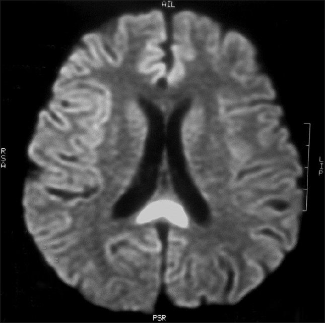
- MRI of brain FLAIR sequence showing hyperintensity of splenium of corpus callosum (the Boomerang sign)

- Left-sided LMN facial nerve palsy
She came for regular follow-ups, was doing well. A repeat MRI of brain was done after 2 months, it showed complete resolution of the splenial hyperintensity in FLAIR [Figure 4].

- Repeat MRI of brain FLAIR sequence showing complete resolution of the hyperintensity of SCC
Discussion
While the incidence of cryptococccal meningitis in the developed world has declined with widespread, early antiretroviral therapy (ART), cryptococcal disease remains a major opportunistic infection and leading cause of mortality in patients infected with HIV in much of the developing world. Most HIV-related cases are caused by Cryptococcus neoformans var. grubii (serotype A), while var. neoformans (serotype D) is responsible for a proportion, especially in Europe, and there are a small number of Cryptococcus gattii infections (formerly C. neoformans serotypes B and C).[12]
HIV-associated cryptococcal meningitis usually presents as a subacute meningo-encephalitis in profoundly immunosupressed patients (CD4 cell counts <100 cells/ml), with malaise, headache, fever and, later, visual disturbance, ataxia, and altered mental status. Signs, if present, may include meningismus, papilloedema, cranial nerve palsies [particularly sixth nerve palsies indicative of raised pressure in cerebrospinal fluid (CSF)] and reduced consciousness level.[3] MRI of brain in cryptococcal meningitis may show cryptococcoma(s), dilated Virchow Robin spaces, etc.
Transient signal abnormality involving solely the splenium of corpus callosum on MRI is not frequently encountered in clinical practice. Two types of lesions have been described, a uniform hyperintensity of the splenium of corpus callosum is called the “Boomerang sign” and a semilunar hyperintensity involving only the posterior part is known as the “Miniboomerang sign”. The occurrence of this abnormality was first described by Chason et al. as a transient post-ictal focal edema denoting transhemispheric propagation of seizure through the corpus callosum.[4] Since then, various etiological factors have been associated with the transient hyperintensities of the splenium [also called as transient splenial lesion (TSL)]. Epilepsy, abrupt change or overdose of antiepileptic drugs, infections like Epstein barr virus, malaria, salmonellosis, pre-eclampsia, acute disseminated encephalomyelitis (ADEM), vitamin B-12 deficiency, multiple sclerosis, herpes simplex encephalitis, trauma, etc., are described in literature by different workers.[56789]
The pathogenesis of the transient changes in the splenium of corpus callosum (SCC) is unknown. In encephalopathy/encephalitis, presumed mechanism was an acute intramyelinic edema due to the separation of myelin layers and leading to transient abnormalities on MRI.[10] Tada et al. proposed that SCC lesions may be secondary to intramyelinic edema from separation of myelin layers, or that an influx of inflammatory cells and macromolecules, combined with cytotoxic edema causes decreased Apparent Diffusion Coefficient levels (ADC). They considered that ADC may return to normal if the cause for its decrease resolved quickly.[11]
Breakdown of the blood-brain barrier (BBB), producing transient focal edema has been implicated by some authors in patients having seizures.[612]
Oster et al. reported two bitemporal lobe epilepsy patients showing a transient SCC lesion, a structure involved in seizure propagation but not seizure initiation. They suggested that the possible mechanism involved a transient disturbance of energy metabolism and ionic transport resulting in reversible myelin vacuolization and intramyelin edema due to excessive repetitive activity of the commissural projection from the temporal structure in these patients.[13] The nerve fiber composition within the splenium of corpus callosum is almost same as other parts of the corpus callosum, but studies have demonstrated different blood supply of the splenium (vertebrobasilar system) from the rest of corpus callosum (carotid system).[141516]
Cryptococcal meningitis has not yet been implicated in the causation of reversible splenial lesions in MRI. The frequently described association of seizures could not be attributed here as she never had convulsions in the past and during the entire course of hospitalisation. The Epstein barr virus-PCR came negative. Although there is a report of splenial hyperintensity with recurrent herpes simplex encephalitis in an infant, the MRI finding of our case can not be attributed to a herpetic etiology due to the lack of other suggestive MRI findings, the CSF report and most importantly, remarkable clinical improvement without intravenous antiviral drugs like acyclovir.[5] We kept the possibility of immune reconstitution inflammatory syndrome (IRIS) in mind, but the CSF features were not matching.
The diagnosis of Cryptococcus meningitis in this case was certain and so this appears to be another cause of the typical vanishing splenium-of-corpus callosum (SCC) lesion in MRI.
We had to start antitubercular drugs initially because of its very high prevalence in this part of the world and because of the delay in the availability of the test results (e.g., CSF EBV-PCR, cryptococcal antigen, etc.) in the hospital we work in.
Conclusions
Transient hyperintensities of splenium of corpus callosum in MRI (the so called Boomerang and Miniboomerang signs) are still uncommon findings in clinical practice. Among the various causes published in literature, persistant seizures have been attributed most commonly. To the best of our knowledge, cryptococcal meningitits has never been described to cause such abnormality. Our idea of reporting this case is to document that such transient MRI findings may also occur in Cryptococcus meningitis along with the other causes previously described and the treating physician must consider all these possibilities once the Boomerang or Miniboomerang sign is seen in MRI of brain, more so if the patient is not an epileptic or is not receiving any antiepileptic drug therapy.
Source of Support: Nil.
Conflict of Interest: None declared.
References
- Cryptococcus gattii infection: Characteristics and epidemiology of cases identified in a South African province with high HIV seroprevalence, 2002-2004. Clin Infect Dis. 2006;43:1077-80.
- [Google Scholar]
- “Cryptococcosis”. In: Longo, Fauci, Kasper, Hauser, Jameson, Loscalzo, eds. In Harrisons Principles of Internal Medicine (18th ed). New York: McGraw-Hill; 2012. p. :1648-51.
- [Google Scholar]
- Transient Splenial Edema in Epilepsy: MR Imaging Evaluation Proceedings of the 34th annual meeting of the American Society of Neuroradiology. 1996 June 21-27
- [Google Scholar]
- MRI findings of recurrent herpes simplex encephalitis in an infant. Pediatr Radiol. 2003;33:725-8.
- [Google Scholar]
- Transient lesion in the splenium of the corpus callosum: Three further cases in epileptic patients and a pathophysiological hypothesis. J Neurol Neurosurg Psychiatry. 2001;70:459-63.
- [Google Scholar]
- Focal lesion in the splenium of the corpus callosum in epileptic patients: Antiepileptic drug toxicity? AJNR Am J Neuroradiol. 1999;20:125-9.
- [Google Scholar]
- Clinical implications of splenium magnetic resonance imaging signal changes. Arch Neurol. 2005;62:433-7.
- [Google Scholar]
- Transient focal lesion in the splenium of the corpus callosum: MR imaging with an attempt to clinical-physiopathological explanation and review of the literature. Radiol Med. 2007;112:921-35.
- [Google Scholar]
- Transient splenial lesion of the corpus callosum in clinically mild influenza associated encephalitis/encephalopathy. AJNR Am J Neuroradiol. 2006;27:1983-6.
- [Google Scholar]
- Clinically mild encephalitis/encephalopathy with a reversible splenial lesion. Neurology. 2004;63:1854-8.
- [Google Scholar]
- Transient postictal magnetic resonance imaging abnormality of the corpus callosum in a patient with epilepsy. Case report and review of the literature. J Neurosurg. 2002;97:714-7.
- [Google Scholar]
- Diffusion-weighted imaging abnormalities in the splenium after seizures. Epilepsia. 2003;44:852-4.
- [Google Scholar]
- The size and fiber composition of the corpus callosum with respect to gender and schizophrenia: A post-mortem study. Biol Psychiatry. 1999;45:1120-7.
- [Google Scholar]
- Vascularisation arte’rielle et veineuse du corps calleux. Neurochirurgie. 1998;4(Suppl 1):31-7.
- [Google Scholar]


