Translate this page into:
Author's reply
This is an open-access article distributed under the terms of the Creative Commons Attribution-Noncommercial-Share Alike 3.0 Unported, which permits unrestricted use, distribution, and reproduction in any medium, provided the original work is properly cited.
This article was originally published by Medknow Publications and was migrated to Scientific Scholar after the change of Publisher.
Sir,
Thank you for giving us an opportunity to reply to the letter “ Acute inter-hemispheric subdural hematoma in a kabaddi player: A comment”.[1] The author has provided very nice plain CT scan images of two separate patients depicting the appearance of straight and sagittal sinus, seen as hyperdense line. It is well known that the normal variations in cerebral venous anatomy are commonly encountered when interpreting imaging studies and may cause diagnostic confusion.[2–4] The great vein of Galen and straight sinus, as they are surrounded by cenebrospinal fluid and brain, are almost always visualized on CT scan particularly if the study is performed with contrast enhancement, with a quite characteristic appearance.[5] As described by the author, an axial section through the tentonium near its apex with the plane of section slightly inclined to the long axis of the straight sinus would include the great vein of Galen and, more posteriorly, would produce a separate elongated configuration.[5] This hyperdense appearance can be due to the partial volume of straight and sagittal sinuses and also we agree with the author that the subdural hematoma will never or rarely have inverted Y-shaped most posterior end (as also true in the present case-Figure 1, small arrow).[6]
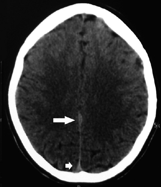
- TCT scan showing the appearance of acute subdural hematoma (large arrow), normal inverted ‘Y’ shaped appearance of the posterior part of the straight sinus (small arrow) and thin uniform hyperdense appearance of the straight sinus (in between arrows).
As shown by the author, the outline of the sinus was quite uniform. However, as can be noted in the reported case, it was irregular and thick particularly at the anterior third part in proximity to the margins of the falx cereberi [Figure 1, large arrow]. Considering the clinical picture and imaging findings, we anticipated that the patient would have sustained injury and diagnosis of acute subdural hematoma was suspected. The usefulness of noninvasive means of evaluating the intracranial venous system including MR venography or CT venography has been increasingly recognized,[78] 3D-CTA can evaluate venous variations of the galenic system and provides useful information.[8] However, because of the non-availability of MRI angiography and venography, these sequences were not performed. Regarding follow up, the patient was doing well at one year follow up. The issues raised by the author are well taken and while working with limited resources, there is a need to interpret data carefully and if feasible one should further investigate the patient with better imaging modalities to confirm the pathology.
References
- Acute inter-hemispheric subdural hematoma in a kabaddi player: A comment. J Neurosci Rural Pract. 2011;2:204-06.
- [Google Scholar]
- Normal and variant anatomy of the dural venous sinuses. Semin Ultrasound CT MR. 1994;15:499-519.
- [Google Scholar]
- Congenital and acquired abnormalities of the dural venous sinuses. Semin Ultrasound CT MR. 1994;15:520-539.
- [Google Scholar]
- Cerebral venous sinuses: Anatomical variants or thrombosis? Acta Anat (Basel). 1988;133:318-24.
- [Google Scholar]
- The Tentorium in Axial Section. I. Normal CT Appearance and Non-neoplastic Pathology. Radiology. 1977;123:631-8.
- [Google Scholar]
- Acute inter-hemispheric subdural hematoma in a Kabaddi player. J Neurosci Rural Pract. 2010;1:122-3.
- [Google Scholar]
- Cerebral MR venography: Normal anatomy and potential diagnostic pitfalls. AJNR Am J Neuroradiol. 2000;21:74-8.
- [Google Scholar]
- Three-dimensional computed tomography angiography of the galenic system for the occipital transtentorial approach. Neurol Med Chir (Tokyo). 2005;45:387-393.
- [Google Scholar]


