
Translate this page into:
A case of a massive mandibular schwannoma
This is an open-access article distributed under the terms of the Creative Commons Attribution-Noncommercial-Share Alike 3.0 Unported, which permits unrestricted use, distribution, and reproduction in any medium, provided the original work is properly cited.
This article was originally published by Medknow Publications and was migrated to Scientific Scholar after the change of Publisher.
Abstract
Schwannoma, a benign nerve sheath tumor is relatively rare in occurrence and even rarer in sites, such as jaw bones. There are only 45 reported cases of intraosseous schwannoma of the jaws reported in the literature. We report a rare case of mandibular schwannoma in a 50-year-old Indian male. The clinical features resembled that of a residual cyst, fibro-osseous lesion or an odontogenic tumor/cyst. Radiological differential diagnoses of ameloblastoma or odontogenic keratocyst was made based on the findings of the orthopantomogram. The lesion was examined histopathologically and a final diagnosis of schwannoma arising from the inferior alveolar nerve was made. The aim of this report is to add information to the existing sparse literature on intraosseous schwannomas of the jaw.
Keywords
Intraosseous
mandible
schwannoma

Introduction
Schwannoma also called as Neurilemmoma is a benign neoplasm of the Schwann cells surrounding the nerves.[1] The first reported literature of schwannoma was in the year 1910 by Verocay.[2] This benign lesion is frequently located in the soft tissues of head and neck region.[34] However, intraosseous schwannoma are extremely rare in jaw bones and represent less than 1% of benign primary tumors of the jaws.[5]
Case Report
A 42-year-old man hailing from Dakshina Kannada district in Karnataka, India, reported to us with a slowly growing swelling on the left side of lower jaw since the previous 4 years. Three years ago teeth in the same region became loose and were extracted at a primary health center. The patient had visited the same health center a number of times because of the swelling. Medications were prescribed each time but there was no decrease in the size of the swelling. The patient was then referred to our institution for further treatment.
On examination, a diffuse swelling was observed on the left lower third of the face extending to the left submandibular region with normal overlying skin. The swelling was firm in consistency with no localized rise in temperature. Intraoral examination revealed bicortical expansion of the edentulous ridge in the same region with reduced buccal sulcus depth. Residual cyst, fibro-osseous lesion, ameloblastoma, dentigerous cyst, and odontogenic keratocyst were listed as the possible differential diagnoses. A mandibular lateral occlusal radiograph was made, which showed buccolingual expansion in the same region and orthopantomogram revealed the presence of large radiolucent area (5 × 7 cm) on the left side of the mandible involving the body and ramus [Figure 1]. The superior border was very poorly demarcated. A sclerotic rim extending 1 cm below the lower border of the mandible was observed. Anteriorly the lesion extended to the distal aspect of premolar and posteriorly to the ramus. The path of the inferior alveolar nerve could not be traced through the lesion. A biopsy was performed from the intraoral site, middle of the mandibular residual alveolar ridge and evaluated histopathologically. It revealed the presence of spindle-shaped cells with elongated nuclei arranged in typical circular (Antoni A) and linear pattern (Antoni B) along with small cyst-like spaces and eosinophilic Verocay bodies [Figure 2]. Based on these histopathologies, it was diagnosed as schwannoma. Since the lesion was massive and only a thin margin of the base was intact, which could lead to pathological fracture, the entire left segment of the mandible was resected barring the condyle. The left side was then reconstructed with bone plate and stabilized with interdental wiring for 3 weeks before being discharged from the hospital. The excised specimen was histopathologically reviewed and the previous diagnosis of schwannoma was confirmed. The patient was followed-up for 6 months during which the patient had of localized numbness on the left side of the lower lip. The patient was also advised prosthetic rehabilitation but failed to keep up with further appointments.

- (Left side) Mandibular lateral occlusal radiograph revealing buccolingual expansion of the body of mandible on the left side. (Right side) Orthopantomogram showing large radiolucent area in the left body and ramus region of mandible
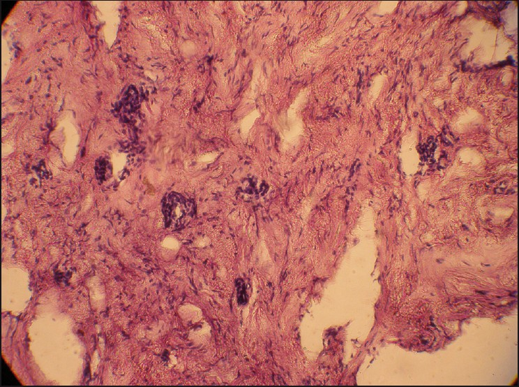
- Photomicrograph (hematoxylin and eosin stain; original magnification ×100) showing cells with elongated spindle-shaped nuclei (Antoni A type) and also homogenous acellular areas (Verocay bodies)
Discussion
Schwannoma also known as neurinoma or neurilemmoma is a benign nerve sheath tumor.[6] Intraoral schwannomas are rare and intrabony occurrence is even rarer.[5] A recent review of literature revealed that only 44 cases of intrabony schwannomas have been reported. Three possible mechanisms of bone involvement by schwannoma have been explained: (1) a tumor may arise within the bone; (2) a tumor may arise within the nutrient canal and produce canal enlargement; or (3) a peripheral tumor causing bone enlargement.[7] In our case a combination of the first 2 mechanisms could be the cause of bone involvement. In most of the reported cases mandible is affected, probably due to the long intrabony course of mandibular nerve.[8] The same nerve was involved in our case on the left side of the mandible. Most common clinical presentation of schwannoma is swelling followed by pain and paresthesia.[9] Swelling was the only feature observed in our patient. Radiographically schwannomas present as well-defined unilocular radiolucency with sclerotic borders but sometimes features, such as cortical expansion, root resorption, spotty calcification, and peripheral scalloping can be evident.[10] Unilocular radiolucency with cortical expansion was observed in our case. Microscopically spindle-shaped cells in Antoni A and Antoni B arrangement interspersed with hyalinized Verocay bodies are characteristic features.[11] Similar histopathological features were noticed in our case. Surgical treatment was carried out in our patient, which is the most widely used method to prevent recurrence.[610]
Conclusion
Intraosseous schwannoma although extremely rare, knowledge of the clinical radiological and histopathological features is extremely important for prompt diagnosis and appropriate management.
Source of Support: Nil
Conflict of Interest: None declared.
References
- Schwannoma with secondary erosion of the mandible: Case report with review of literature. Dentomaxillofac Radiol. 2006;35:456-60.
- [Google Scholar]
- Atypical central neurilemmoma of the mandible. Oral Surg Oral Med Oral Pathol. 1984;59:275-8.
- [Google Scholar]
- Central neurilemmoma of maxilla. Oral Surg Oral Med Oral Pathol Oral Radiol Endod. 1995;79:41-3.
- [Google Scholar]
- Neurilemmoma and related tumors. In: Unni KK, ed. . Dahlin's bone tumors: General aspects and data on 11,087 cases (5th ed). Philadelphia: Lippincott-Raven; 1996. p. :343-347.
- [Google Scholar]
- Solitary intraosseous neurilemmoma of the tibia: Review of intraosseous neurilemmoma and neurofibromas. Clin Orthop. 1976;117:271-81.
- [Google Scholar]
- Intrabony neurilemmoma: Diagnosis and management. J Am Dent Assoc. 1998;129:729-32.
- [Google Scholar]
- Intraosseous schwannoma of the mandible: A case report and review of literature. Oral Surg Oral Med Oral Pathol Oral Radiol Endod. 2003;96:54-65.
- [Google Scholar]
- Intraoral Schwannoma of mandibular symphysis: Case report. Braz Dent J. 2006;17:255-8.
- [Google Scholar]


