
Translate this page into:
Inca - interparietal bones in neurocranium of human skulls in central India
This is an open-access article distributed under the terms of the Creative Commons Attribution-Noncommercial-Share Alike 3.0 Unported, which permits unrestricted use, distribution, and reproduction in any medium, provided the original work is properly cited.
This article was originally published by Medknow Publications and was migrated to Scientific Scholar after the change of Publisher.
Abstract
Inca bones are accessory bones found in neurocranium of human skulls. Occurrence of Inca bones is rare as compared to other inter sutural bones such as wormian bones. These Inca ossicles are regarded as variants of the normal. The reporting of such occurrences is inadequate from Central India.
Objectives:
To find the incidence of Inca variants in Central India.
Materials and Methods:
In the present study, 380 dried adult human skulls were examined. All specimen samples were procured from various Medical colleges of Central India. They were analyzed for gross incidence, sexual dimorphism and number of fragments of Inca bones.
Results:
Gross incidence of Inca bones was found to be 1.315 %. Incidence rate was higher in male skulls than female skulls (male: 1.428%; female: 1.176%). The Inca bones frequently occurred signally. Out of the five observed Inca ossicles, two were fragmented.
Conclusions:
This data gives idea regarding gross incidence, sexual dimorphism and number of fragments of Inca bones in neurocranium of human skulls from Central India. The knowledge of this variable is useful for neurosurgeons, anthropologists and radiologists.
Keywords
Neurocranium
sutural bones
inca bones
central India

Introduction
The human skull is made up of 22 paired and unpaired ossicles, which are classified as viscerocranium and neurocranium. The neurocranial bones of variable size and shape are often encountered at the fontanellae or along the sutures. Inca ossicles are found in the line of the interparietal suture. Inca ossicles are bounded by lamdoid suture and sutura mendosa. They were previously known as os-Incae, os-ipactal, Goethe's ossicles.[1] Gordan, 1963, describing Inca said it resembles a triangular architectural monument design of the Inca tribe.[2] Inca bones were supposed to be present in the Inca tribals in South Andes – America 1200-1597 A.D. The members of Royal family of Inca tribe had crown-like configuration on their head.[3] Hence, thereafter, these ossicles have been known as Inca. The occipital bones ossify in four centers; one for membranous squamous part, one for the basal cartilaginous part and two for the condylar part of the occipital bone.[4] Inter-parietal bones may occur due to failure of fusion between primary and secondary centers of ossification of occipital bone.[5]
The occurrence of Inca variable is rare. The skull bone fractures in infancy and early childhood may associate with large intersutural bones. These Inca bones may give a false appearance of fracture on Roentgenographs. Such bones may produce complications during burr-hole surgeries and their extensions may lead to continuation of fracture lines. Due to clinical implication information of presence of Inca bones, their incidence, sexual dimorphism and number of fragments is essential to clinicians. Many researchers have reported Inca and other neurocranial variables throughout the world.[67] The presence of Inca osssicles may be used as corroborative evidence for familial identity in medico legal cases.[8] Data on incidence, sexual dimorphism and number of fragments of Inca bones from Central India is underreported. Hence, this study was undertaken to find the incidence of this variant in the Central India.
Materials and Methods
The study was conducted at the Department of Anatomy of a tertiary care hospital and medical college of Central India during 1992 to 1994. A total of 380 dried adult human skulls were examined for the study. All the samples were procured from various medical institutes in Central India. All selected samples were without any signs of ante-mortem or post-mortem injuries. They were analyzed for the gross incidence, sexual dimorphisms and number of fragments of Inca ossicles. The collected data was analyzed statistically.
Results
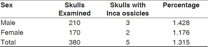
Gross incidence of Inca ossicles was 1.315 % (5 Inca bones in 380 skulls) [Figure 1, Table 1]. Sexual dimorphism for the presence of Inca bones was observed. Incidence rate was higher in male skulls than female skulls (male: 1.428%; female: 1.176%) [Table 1]. Out of the five observed Inca ossicles, two were fragmented.
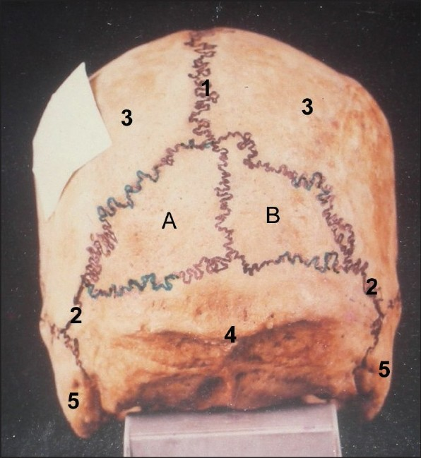
- Photograph showing large Inca ossicles. A, B: Inca ossicles, 1: Interparietal suture, 2: Parietooccipital suture, 3: Parietal bone, 4: Occipital bone, 5: Mastoid process
Discussions
Inca ossicles are accessory bones found in human skulls as interparital bones. Gross incidence of Inca ossicles was 1.315 %. A very high incidence (27.71 %) of Inca ossicles was found in the pre-hispanic skulls dated between 300-1200 A.C.[9] Berry et al. reported 2.9 to 4.6 % incidence in American population of South West coast, which is higher than that of present study.[6] Tsunehiko et al. also reported low incidence of Inca ossicles in Asians as compared to new world Population.[10] Reported a different incidence in population of North India (0.4%).[11] These findings might be indicative of natural evolutionary changes, probably swinging on either side of the spectrum.
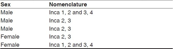
The present study reports an incidence of higher in male (male: 1.428% and female: 1.176 %) for the female skull, which was in concurrence with the incidence rates of Berry and berry (Male: 4.6 % and Female: 2.9%).[6] In the present study, maximum of two fragments of Inca bones were observed in three skulls. Purkait and Chandra classified Inca ossicles on a developmental basis.[8] They numbered the four ossification centers from left to right as 1, 2, 3 and 4, out of which 1 and 4 represented lateral ossification centers while element 2 and 3 represented the medial ossification centers. The respective contributing component is indicated with their numbers prefixed by the term Inca, e.g. if Inca observed in adult skull is contributed by nonfused medial pairs of centers, then it is known as Inca 2 and 3; if the centers were fused, the so formed Inca is termed Inca 2, 3. The nomenclature of Inca ossicles as per Purkait- Chandra classification found in the present study is indicated in Table 2.
The occurrence of Inca ossicles in skulls may be explained on the basis of embryological and evolutionary development of skull. The chondrocramium of fish skull and its components are forerunners of human skull base. These primitive components can be distinguished in the genesis of human skull.[12] This ossification center appeared in fetal skulls in 2nd and 3rd months of intrauterine life.[13] The appearance of additional centers for ossification may lead to occurrence of Inca bones.
Conclusions
Meticulous knowledge regarding gross incidence, sexual dimorphism and number of Inca ossicles in human skulls in Central India may be useful to neurosurgeons, orthopedic surgeons, anthropologists and radiologists.
Source of Support: Nil
Conflict of Interest: None declared.
References
- The skull. In: In. Gray's Anatomy (38th edi). London: Churchill Livingstone; 1995. p. :583-606.
- [Google Scholar]
- The Book of knowledge (6th ed). London: Waverlay Book Co. Ltd; 1963. p. :161-6.
- The Incas and Their Ancestors. In: In. The Archaeology of Peru. London: Thames and Hudson; 1992. p. :8.
- [Google Scholar]
- External Skull. In: In. Gray's Anatomy (40th ed). London: Churchill Livingstone; 2008. p. :411-8.
- [Google Scholar]
- Developmental studies on the interparietal part of the human occipital squama. J Anat. 1993;182:197-204.
- [Google Scholar]
- The identity of Inca, medicolegal importance and suggested nomenclature of variants. J Anat Soc Ind. 1989;38:162-71.
- [Google Scholar]
- Frequency of interparietal bone or Inca bone in pre-Hispanic atacamenos skulls of the north of Chile. Int J Morph 2008 in press
- [Google Scholar]
- Os Incae: Variation in frequency in major human population groups. J Anat. 2001;198:137-52.
- [Google Scholar]
- Incidence of interparietal bones in adult skulls of Agra Region. Anat Anz. 1979;145:528-31.
- [Google Scholar]
- Development of head and neck. In: Human Embryology (3rd ed). Pennsylvania: Churchill Livingstone; 2001. p. :355-78.
- [Google Scholar]
- Der Stirnfortsatze der Schalafenschuppe bei den Primaten. S Math Phys Cl Ak Wiss Zu Mun. 1898;27:227-70.
- [Google Scholar]


